

Role of malignant ascites on human mesothelial cells and their gene expression profiles
- PMID: 24761768
- PMCID: PMC4008408
- DOI: 10.1186/1471-2407-14-288
Role of malignant ascites on human mesothelial cells and their gene expression profiles
Abstract
Background: Malignant ascites is often present at diagnostic in women with advanced ovarian cancer (OC) and its presence is associated with a worse outcome. Human peritoneal mesothelial cells (HPMCs) are key components of malignant ascites. Although the interplay between HPMCs and OC cells is believed to be critical for tumor progression, it has not been well characterized. The purpose of this study was to assess the effect of ascites on HPMCs and clarify the role of HPMCs in OC progression.
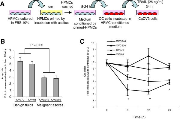
Methods: Human OC ascites and benign peritoneal fluids were assessed for their ability to stimulate HPMC proliferation. Conditioned medium from ascites- and benign fluid-stimulated HPMCs were compared for their ability to attenuate apoptosis induced by TNF-related apoptosis-inducing ligand (TRAIL). We conducted a comparative analysis of global expression changes in ascites-stimulated HPMCs using Agilent oligonucleotide microarrays.
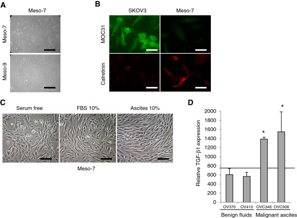
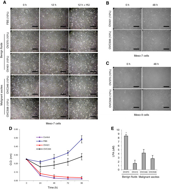
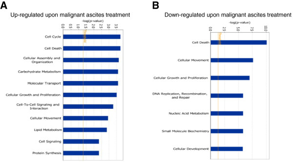
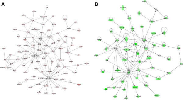
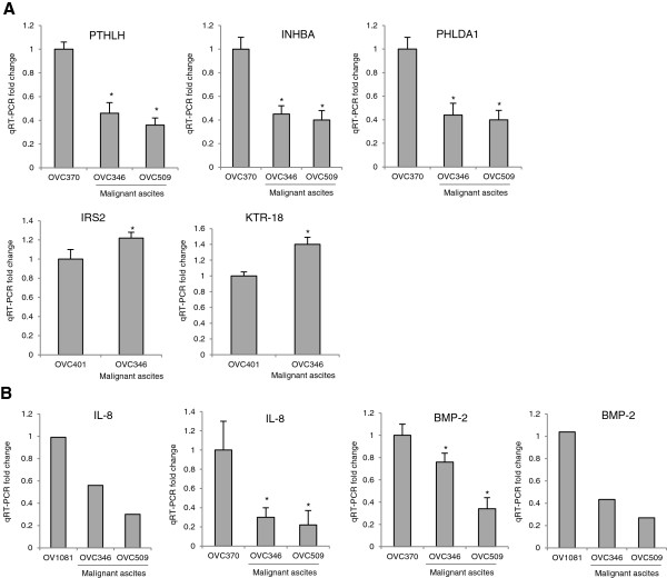
Results: As compared to benign peritoneal fluids, malignant ascites stimulated the proliferation of HPMCs. TRAIL-induced apoptosis was attenuated in OC cells exposed to conditioned medium from ascites-stimulated HPMCs as compared to OC cells exposed to conditioned medium from benign fluid-stimulated HPMCs. A total of 649 genes were differentially expressed in ascites-stimulated HPMCs. Based on a ratio of more than 1.5-fold and a P < 0.05, 484 genes were up-regulated and 165 genes were down-regulated in ascites-exposed HPMCs. Stimulation of HPMCs with OC ascites resulted in differential expression of genes mainly associated with the regulation of cell growth and proliferation, cell death, cell cycle and cell assembly and organization, compared to benign peritoneal fluids. Top networks up-regulated by OC ascites included Akt and NF-κB survival pathways whereas vascular endothelial growth factor (VEGF) pathway was down-regulated.
Conclusions: The results of this study not only provide evidence supporting the importance of the interplay between cancer cells and HPMCs but also define the role that the tumor environment plays in these interactions.
Figures
Similar articles
-
Ovarian cancer-derived ascitic fluids induce a senescence-dependent pro-cancerogenic phenotype in normal peritoneal mesothelial cells.Cell Oncol (Dordr). 2016 Oct;39(5):473-481. doi: 10.1007/s13402-016-0289-1. Epub 2016 Jul 21. Cell Oncol (Dordr). 2016. PMID: 27444787
-
Ascites from ovarian cancer patients stimulates MUC16 mucin expression and secretion in human peritoneal mesothelial cells through an Akt-dependent pathway.BMC Cancer. 2019 Apr 30;19(1):406. doi: 10.1186/s12885-019-5611-7. BMC Cancer. 2019. PMID: 31039761 Free PMC article.
-
Ovarian cancer ascites enhance the migration of patient-derived peritoneal mesothelial cells via cMet pathway through HGF-dependent and -independent mechanisms.Int J Cancer. 2015 Jul 15;137(2):289-98. doi: 10.1002/ijc.29385. Epub 2014 Dec 18. Int J Cancer. 2015. PMID: 25482018
-
Immune Tumor Microenvironment in Ovarian Cancer Ascites.Int J Mol Sci. 2022 Sep 14;23(18):10692. doi: 10.3390/ijms231810692. Int J Mol Sci. 2022. PMID: 36142615 Free PMC article. Review.
-
Spheroid Formation and Peritoneal Metastasis in Ovarian Cancer: The Role of Stromal and Immune Components.Int J Mol Sci. 2022 Jun 1;23(11):6215. doi: 10.3390/ijms23116215. Int J Mol Sci. 2022. PMID: 35682890 Free PMC article. Review.
Cited by
-
Cancer-associated mesothelial cell-derived ANGPTL4 and STC1 promote the early steps of ovarian cancer metastasis.JCI Insight. 2023 Mar 22;8(6):e163019. doi: 10.1172/jci.insight.163019. JCI Insight. 2023. PMID: 36795484 Free PMC article.
-
Regulation of Mesothelial Cell Fate during Development and Human Diseases.Int J Mol Sci. 2022 Oct 8;23(19):11960. doi: 10.3390/ijms231911960. Int J Mol Sci. 2022. PMID: 36233262 Free PMC article. Review.
-
Ovarian cancer-derived ascitic fluids induce a senescence-dependent pro-cancerogenic phenotype in normal peritoneal mesothelial cells.Cell Oncol (Dordr). 2016 Oct;39(5):473-481. doi: 10.1007/s13402-016-0289-1. Epub 2016 Jul 21. Cell Oncol (Dordr). 2016. PMID: 27444787
-
Molecular mediators of peritoneal metastasis in pancreatic cancer.Cancer Metastasis Rev. 2020 Dec;39(4):1223-1243. doi: 10.1007/s10555-020-09924-4. Epub 2020 Aug 11. Cancer Metastasis Rev. 2020. PMID: 32780245 Free PMC article. Review.
-
Mesothelial cells interact with tumor cells for the formation of ovarian cancer multicellular spheroids in peritoneal effusions.Clin Exp Metastasis. 2016 Dec;33(8):839-852. doi: 10.1007/s10585-016-9821-y. Epub 2016 Sep 9. Clin Exp Metastasis. 2016. PMID: 27612856
References
Publication types
MeSH terms
Substances
Grants and funding
LinkOut - more resources
Full Text Sources
Other Literature Sources
Medical
Molecular Biology Databases
Research Materials
