Review
. 2014 Aug;40(5):520-43.
doi: 10.1111/nan.12150.

Review: Hippocampal sclerosis in epilepsy: a neuropathology review

Affiliations
Free PMC article
Review

Review: Hippocampal sclerosis in epilepsy: a neuropathology review

Maria Thom. Neuropathol Appl Neurobiol. 2014 Aug.
Free PMC article

Abstract

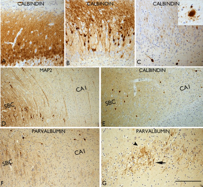
Hippocampal sclerosis (HS) is a common pathology encountered in mesial temporal lobe epilepsy (MTLE) as well as other epilepsy syndromes and in both surgical and post-mortem practice. The 2013 International League Against Epilepsy (ILAE) classification segregates HS into typical (type 1) and atypical (type 2 and 3) groups, based on the histological patterns of subfield neuronal loss and gliosis. In addition, granule cell reorganization and alterations of interneuronal populations, neuropeptide fibre networks and mossy fibre sprouting are distinctive features of HS associated with epilepsies; they can be useful diagnostic aids to discriminate from other causes of HS, as well as highlighting potential mechanisms of hippocampal epileptogenesis. The cause of HS remains elusive and may be multifactorial; the contribution of febrile seizures, genetic susceptibility, inflammatory and neurodevelopmental factors are discussed. Post-mortem based research in HS, as an addition to studies on surgical samples, has the added advantage of enabling the study of the wider network changes associated with HS, the long-term effects of epilepsy on the pathology and associated comorbidities. It is likely that HS is heterogeneous in aspects of its cause, epileptogenetic mechanisms, network alterations and response to medical and surgical treatments. Future neuropathological studies will contribute to better recognition and understanding of these clinical and patho-aetiological subtypes of HS.

Keywords: hippocampal sclerosis; neuropathology; temporal lobe epilepsy.


Figures

Figure 1
Figure 1
Typical pathology features of hippocampal sclerosis in epilepsy. (A) Luxol fast blue/cresyl violet-stained preparation of a surgical sample with subfield neuronal loss involving CA1 and CA4. (B) GFAP preparation confirming dense fibrillary gliosis in CA1 with a sharp cut-off point (arrows) with the adjacent subiculum (SBC) and (C) higher magnification detail of the abrupt transition of gliosis at the CA1/subiculum (arrows). (D) Granule cell dispersion as seen with cresyl violet and (E) with NeuN showing clusters of dispersed granule cells (inset shows reelin-positive interneurones and bipolar cells in the molecular layer). (F) Neurofilament-positive neurones with enlarged cell bodies in CA4 of the sclerotic hippocampus. (G) CA1 pyramidal cell layer in a patient with MTLE but no evidence of neuronal loss, (H) CA1 with evidence of partial neuronal loss from mid zone and, (I) CA1 with severe neuronal depletion and collapse of the layer. (J) Section of the pes hippocampus from a patients with confirmed classical ILAE type 1 sclerosis in the body; in the pes neuronal loss is limited to the endplate in this case as an illustration of variability in the pattern of atrophy that may occur along the longitudinal hippocampal axis. Bar is equivalent to approximately 1 mm (A, B, J), 250 micrometers (C, F, G, H, I) and 100 micrometers (D, E).
Figure 2
Figure 2
Axonal sprouting in hippocampal sclerosis (HS)/temporal lobe epilepsy (TLE). Mossy fibre sprouting identified with Timm staining in hippocampal sclerosis (HS) (A–C). In (A) the black silver granules are mainly confined to the subgranular zone and significant sprouting into the molecular layer (arrow) is not observed. In (B) there is focal sprouting in the molecular layer (arrow) and in (C) marked sprouting is shown with a dense band of zinc positive granules in the molecular layer. (D) Similar mossy fibre sprouting confirmed with dynorphin immunohistochemistry. (E) Extensive sprouting of neuropeptide Y-positive fibres is seen in the dentate gyrus compared with normal pattern (inset). (F) Sprouting of galanin fibres is shown in the gliotic dentate gyrus from a case with TLE and hippocampal sclerosis. In HS more intense synaptic pattern of staining was noted in the molecular layer of the dentate gyrus in a series compared with mTLE cases without HS (M. Thom, unpub. obs.). (G) The myeloarchitecture can appear abnormal in HS with a random arrangement of fibres permeating the dentate gyrus. (H) Calretinin immunohistochemistry with sprouting fibres through the molecular layer compared with the compact alignment of fibres embracing the dentate gyrus in the normal hippocampus (shown in inset). (I) Calbindin immunohistochemistry with sprouted fibres through the molecular layer in HS and loss of the normal expression in the granule cells. In all images the molecular layer is at the top of each figure and the subgranular zone at the bottom. Bar is equivalent to approximately 50 μm (A, B, C) and 100 μm in others.
Figure 3
Figure 3
Interneuronal changes in hippocampal sclerosis in epilepsy (HS). (A) Normal calbindin pattern with labelling of the granule cells and apical dendrites. (B) Absent or loss of calbindin expression is noted in the basal cells while the dispersed cells are calbindin positive; this is a common pattern observed in HS with granule cell dispersion. (C) Virtual total loss of calbindin expression in all the granule cells; residual hilar calbindin-positive neurones as well as in CA1 (inset) can appear hypertrophic and dysmorphic with abnormal processes extending from the cell body. (D) The sharp transition between the neuronal loss in CA1 and the preserved subicular (SBC) neurones is illustrated with MAP2 staining. (E) Calbindin labelling in the same case as (D) shows relative loss of interneurones and their arborizations in the CA1 as with parvalbumin (F). Increased perisomatic labelling and terminals in the dentate gyrus, as with parvalbumin in an HS case. (G) Complex parvalbumin terminals are seen in the dentate gyrus granule cell layer in HS. Bar is equivalent to approximately 75 μm.
Figure 4
Figure 4
Pathological alterations potentially contributing to epileptogenesis in hippocampal sclerosis (HS). (A) Prominent labelling of voltage gate potassium channel (KV1.1) in residual neurones in CA1 in hippocampal sclerosis; differences were noted in the patterns of expression between hippocampal subfields in HS compared with controls (M. Thom, unpub. obs.). (B) Prominent labelling of cannabinoid receptor is noted in the plexus in the molecular layer of the dentate gyrus in HS. (C) Intense labelling of CA1 neurones with cation-chloride cotransporter (NKCC) in HS. (D) In some cases of HS in epilepsy, particularly in adult onset cases or in the context of recent encephalitis, striking reactive and ‘balloon cell’ gliosis can be seen in the granule cell layer with a proportion of these cells showing membranous CD34 staining, mimicking a focal cortical dysplasia (inset). (E) Immunolabelling with isoform GFAP-delta highlights prominent numbers of small astroglia, particularly in the subgranular zone, in HS. (F) Immunostaining for gap junction protein connexin 43 (Cx43) demonstrated prominent labelling of astrocytic cells in the subgranular zone and occasionally in the molecular layer in a post-mortem HS case. (G) Expression of drug transporter protein, p-glycoprotein, is shown on capillaries within hippocampus. (H) Albumin staining demonstrating focal leakage from small capillary vessels in a case of HS/TLE. (I) Coexpression of vascular endothelial growth factor (VEGF) and hypoxic inducible factor 1 alpha (HIF1-alpha) is shown in pyramidal neurones in HS. Bar is equivalent to approximately 35 μm (A, B, D, E, I); 60 μm (C, E, F, H).
Figure 5
Figure 5
Variability of hippocampal sclerosis (HS) in post-mortem and surgical samples. (A) Surgical hippocampectomy specimens, which on histological examination correlated to (i) end folium gliosis with no evidence of sclerosis; (ii) ILAE Type 3 HS (end-folium sclerosis); (iii) ILAE type 2 HS (CA1 predominant sclerosis); and (iv) ILAE type 1 HS (classical hippocampal sclerosis). In all of the images the arrow indicates the pyramidal cell layer of CA1. (B) A 9.4T MRI image of a post-mortem hippocampus from a patient with longstanding epilepsy and ILAE type 2 (CA1 predominant pattern) of HS at this level (shown in C in a luxol fast blue/cresyl violet preparation), although in other levels the pattern was type 1 (classical). The MRI has the ability to identify subfields and white matter tracts and, indistinctly (arrowed), the dentate gyrus. Improved high fields sequences in the future may be able to identify and define patterns of HS pre-operatively. The MRI image was provided with courtesy of Dr Sofia Eriksson at the Department of Clinical and Experimental Epilepsy, UCL, Institute of Neurology. (D to I) Paired sections of hippocampus from one hemisphere labelled with (D to F) GFAP/counterstained with cresyl violet and (G to I) calretinin: at the level of the subthalamic nucleus (STNc; D, G), lateral geniculate nucleus (LGNc; E, H) and hippocampal tail (F, I). Classical pattern (ILAE type 1) HS is seen in the anterior levels with sprouting of calretinin-positive fibres visible in the dentate gyrus (arrow) at this low magnification; in the tail gliosis and neuronal loss is visible in the CA4 region of the hippocampal tail. Bar is equivalent to approximately 3 mm (D to I).

Similar articles

Cited by

References

    1. Bouchet C, Cazauvieilh CA. De l'épilepsie considerée dans ses rapports avec l'aliénation mentale. Recherche sur la nature et le siège de ces deux maladies. Arch Gen Med. 1825:510–542.
    1. Sommer W. Erkrankung desAmmonshornes als aetiologisches Moment der Epilepsie. Arch Psychiatr Nervenkr. 1880:361–375.
    1. Thom M. Hippocampal sclerosis: progress since Sommer. Brain Pathol. 2009;19:565–572. - PMC - PubMed
    1. Blumcke I. Neuropathology of focal epilepsies: a critical review. Epilepsy Behav. 2009;15:34–39. - PubMed
    1. Blumcke I, Coras R, Miyata H, Ozkara C. Defining clinico-neuropathological subtypes of mesial temporal lobe epilepsy with hippocampal sclerosis. Brain Pathol. 2012;22:402–411. - PMC - PubMed

Publication types