Review
. 2014 Jul;25(7):1387-400.
doi: 10.1681/ASN.2014010117. Epub 2014 Apr 24.

Beyond tissue injury-damage-associated molecular patterns, toll-like receptors, and inflammasomes also drive regeneration and fibrosis

Affiliations
Review

Beyond tissue injury-damage-associated molecular patterns, toll-like receptors, and inflammasomes also drive regeneration and fibrosis

Hans-Joachim Anders et al. J Am Soc Nephrol. 2014 Jul.

Abstract

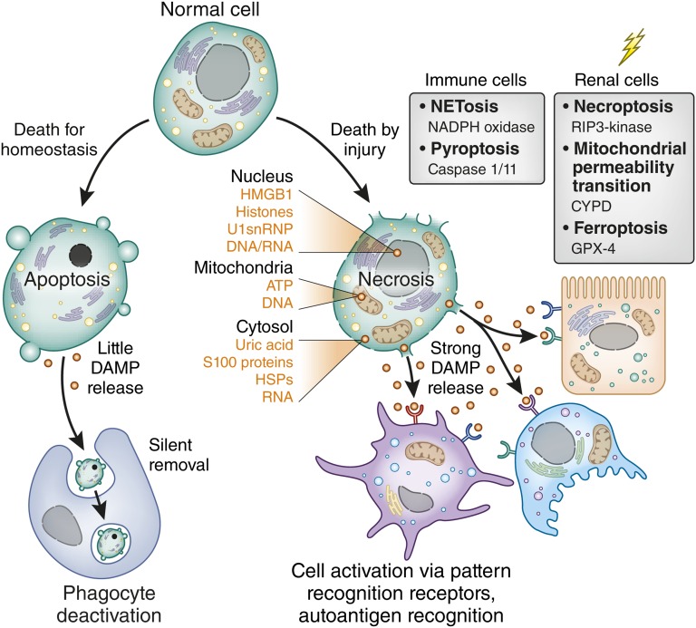
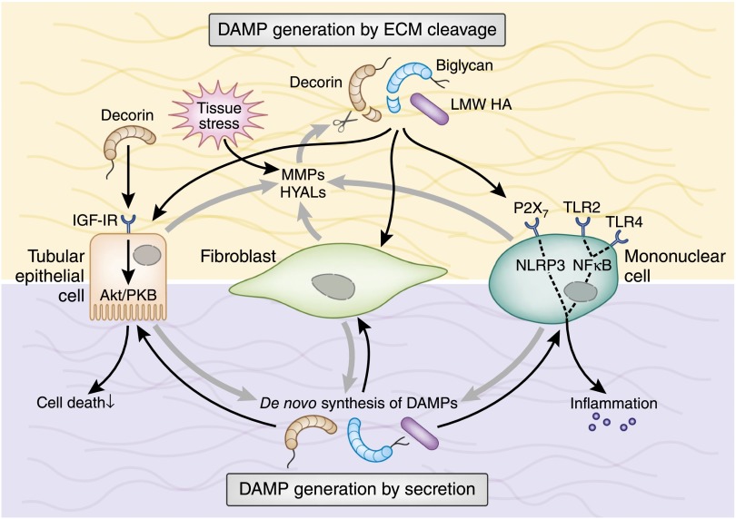
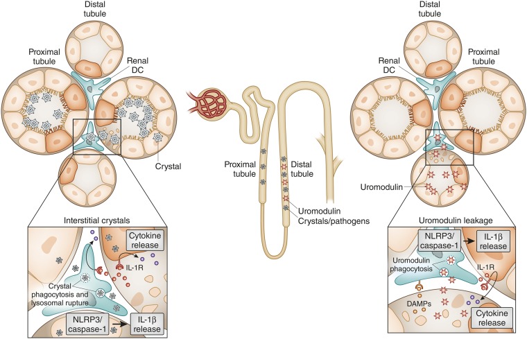
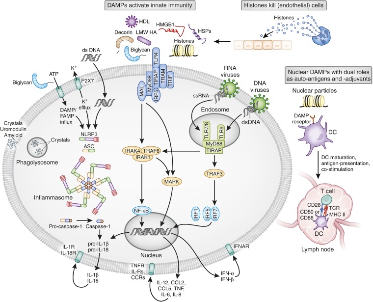
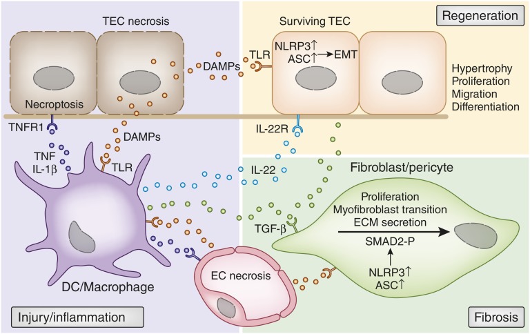
Tissue injury initiates an inflammatory response through the actions of immunostimulatory molecules referred to as damage-associated molecular patterns (DAMPs). DAMPs encompass a group of heterogenous molecules, including intracellular molecules released during cell necrosis and molecules involved in extracellular matrix remodeling such as hyaluronan, biglycan, and fibronectin. Kidney-specific DAMPs include crystals and uromodulin released by renal tubular damage. DAMPs trigger innate immunity by activating Toll-like receptors, purinergic receptors, or the NLRP3 inflammasome. However, recent evidence revealed that DAMPs also trigger re-epithelialization upon kidney injury and contribute to epithelial-mesenchymal transition and, potentially, to myofibroblast differentiation and proliferation. Thus, these discoveries suggest that DAMPs drive not only immune injury but also kidney regeneration and renal scarring. Here, we review the data from these studies and discuss the increasingly complex connection between DAMPs and kidney diseases.

Keywords: ARF; GN; immunology and pathology.


Figures

Figure 1.
Figure 1.
The type of cell death defines DAMP release. Most cells of the body undergo periodic replacement by renewal just like the blood cells. In addition, lymphocyte maturation involves induced cell death to sort out cells with autoreactivity. All such cells undergo apoptosis, a programmed form of cell death that maintains inner and outer membranes to avoid DAMP release. By contrast, cell death induced by injury leads to necrosis. Because of the numerous types of injury (genotoxic stress, toxins, cytokines, oxidative stress), several pathways exist to induce programmed necrosis. Some seem to occur only in specific cell types such as neutrophils (N) or macrophages (MØ). The graph lists essential signaling molecules that trigger this form of cell death. Necrosis implies the disruption of inner and outer membranes, which leads to the release of intracellular DAMPs from various compartments as listed on the right. U1snRNP, U1 small nuclear ribonucleoprotein; NADPH, NAD phosphate dehydrogenase; CYPD, cyclophilin D, GPX-4, glutathione peroxidase 4; HSP, heat shock protein.
Figure 2.
Figure 2.
ECM-related DAMPs are secreted or originate from ECM cleavage. Under tissue stress or injury, resident cells (e.g., TECs and fibroblasts) release MMPs and HYALs to cleave ECM components. Soluble biglycan and decorin and low molecular weight HA act as extracellular DAMPs by interacting with TLR2/TLR4 on the surface of infiltrating mononuclear and resident renal cells. In mononuclear cells, this leads to the activation of NF-κB and production of proinflammatory cytokines and chemokines that, in turn, recruit additional mononuclear cells to the side of injury. Moreover, soluble biglycan and low molecular weight HA activate the NLRP3 inflammasome. Proinflammatory cytokines stimulate mononuclear and renal resident cells to de novo produce DAMPs, thereby creating a positive feedback loop that amplifies the inflammatory response. Besides acting as a DAMP, decorin signals via IGF-IR/Akt in TECs and protects them from apoptosis. LMW, low molecular weight; MMP, matrix metalloproteinase; HYAL, hyaluronidase; IGF-IR, IGF type 1 receptor; PKB, protein kinase B.
Figure 3.
Figure 3.
Crystals and uromodulin act as DAMPs to induce renal inflammation. Crystals precipitate in the proximal tubule, the distal tubule, and/or in the interstitial compartment of the kidney. Crystals kill TECs. In addition, crystals can be taken up by interstitial dendritic cells via phagocytosis. Lysosomal leakage and potassium efflux (not shown) provide a signal to activate the NLRP3 inflammasome, which cleaves caspase-1 and subsequently pro-IL-1β and pro-IL-18 (not shown). IL-1β ligates the IL-1 receptor (IL-1R) on renal parenchymal cells as well as immune cells, which triggers NF-κB–dependent cytokine and chemokine release. In distal tubule injury, uromodulin leakage into the interstitial compartment activates dendritic cells via TLR4 and the NLRP3 inflammasome. As uromodulin also binds to crystals, crystal precipitation in the distal tubule is likely to involve both mechanisms.
Figure 4.
Figure 4.
DAMP effects in immunopathology. DAMPs activate several classes of pattern recognition receptors, which all induce an immediate activation of innate immunity (i.e., systemic and tissue inflammation). Most DAMPs activate TLR2 and TLR4 at the cell surface. In particular, particles enter the cells via phagocytosis and trigger assembly and activation of the NLRP3 inflammasome. Nucleic acid–related DAMPs activate TLR7 and TLR9 in intracellular endosomes. All pattern recognition receptors finally drive the secretion of proinflammatory cytokines that then activate cytokine receptors on the same cell or on other cells. Extracellular histones also directly kill (endothelial) cells. The molecular mechanism of this process is poorly defined but may involve surface charge. Certain nuclear DAMPs also act as autoantigens in systemic lupus and contribute to lupus nephritis. Their adjuvant-like ability to also activate antigen-presenting cells via TLR7 and TLR9 strongly promotes autoimmunization and the expansion of autoreactive lymphocytes. Local release within the kidney also promotes intrarenal autoantigen recognition (e.g., by circulating autoantibodies and subsequent immune complex GN; not shown). LMW, low molecular weight; HSP, heat shock protein; MAL, MyD88 adaptor–like; IRF, IFN regulatory factor; TRAP, TNF receptor–associated protein; TRAF, TNF receptor–associated factor; TRIF, TIR domain–containing adapter inducing IFN-β; ssRNA, single-stranded RNA; dsDNA, double-stranded RNA; DC, dendritic cell;ASC, apoptotic speck protein; IRAK, IL-1 receptor–associated kinase; TIRAP, Toll IL-1 receptor domain–containing adaptor protein; TCR, T-cell receptor; IFNAR, IFN-α receptor; IL-R, IL receptor; CCR, CC-chemokine receptor.
Figure 5.
Figure 5.
How DAMPs trigger immune injury, regeneration and fibrosis. Early injury-induced necrosis leads to DAMP release by renal endothelial and epithelial cells. These DAMPs activate pattern recognition receptors such as TLRs or inflammasomes in intrarenal mononuclear phagocytes such as dendritic cells (DCs) or macrophages. Upon activation, these cells produce inflammatory cytokines and chemokines that cause further renal cell necrosis, (e.g., by TNF-induced necroptosis). This autoamplification loop of injury and inflammation is further accelerated by chemokine-driven leukocyte influx (not shown). DAMP release by necrotic cells also triggers regenerative mechanisms, directly and indirectly. Certain DAMPs activate TLR2 on renal progenitor cells, which accelerates tubular repair. In addition, TLR4 activation of renal dendritic cells triggers IL-22 release, which specifically activates the IL-22 receptor on TECs and accelerates tubular re-epithelialization. DAMPs also trigger fibrosis by activating pericytes, fibroblasts, and mesangial cells (latter not shown). TLR activation induces NLRP3 and ASC expression, which are needed for SMAD2 phosphorylation as a critical step in TGF-β receptor signaling. This was DAMPs drive the transition into myofibroblasts, proliferation, and ECM secretion. The same process also triggers TGF-β receptor–dependent epithelial-mesenchymal transition of renal epithelial cells. ASC, apoptotic speck protein; EMT, epithelial-mesenchymal transition; EC, endothelial cell.

Similar articles

Cited by

References

    1. Wallach D, Kang TB, Kovalenko A: Concepts of tissue injury and cell death in inflammation: A historical perspective. Nat Rev Immunol 14: 51–59, 2014 - PubMed
    1. Medzhitov R: Origin and physiological roles of inflammation. Nature 454: 428–435, 2008 - PubMed
    1. Matzinger P: Tolerance, danger, and the extended family. Annu Rev Immunol 12: 991–1045, 1994 - PubMed
    1. Kurts C, Panzer U, Anders HJ, Rees AJ: The immune system and kidney disease: Basic concepts and clinical implications. Nat Rev Immunol 13: 738–753, 2013 - PubMed
    1. Leemans JC, Stokman G, Claessen N, Rouschop KM, Teske GJ, Kirschning CJ, Akira S, van der Poll T, Weening JJ, Florquin S: Renal-associated TLR2 mediates ischemia/reperfusion injury in the kidney. J Clin Invest 115: 2894–2903, 2005 - PMC - PubMed

Publication types