. 2014 Apr 24;10(4):e1004291.
doi: 10.1371/journal.pgen.1004291. eCollection 2014 Apr.

A quality control mechanism coordinates meiotic prophase events to promote crossover assurance

Affiliations

A quality control mechanism coordinates meiotic prophase events to promote crossover assurance

Alison J Deshong et al. PLoS Genet. .

Abstract

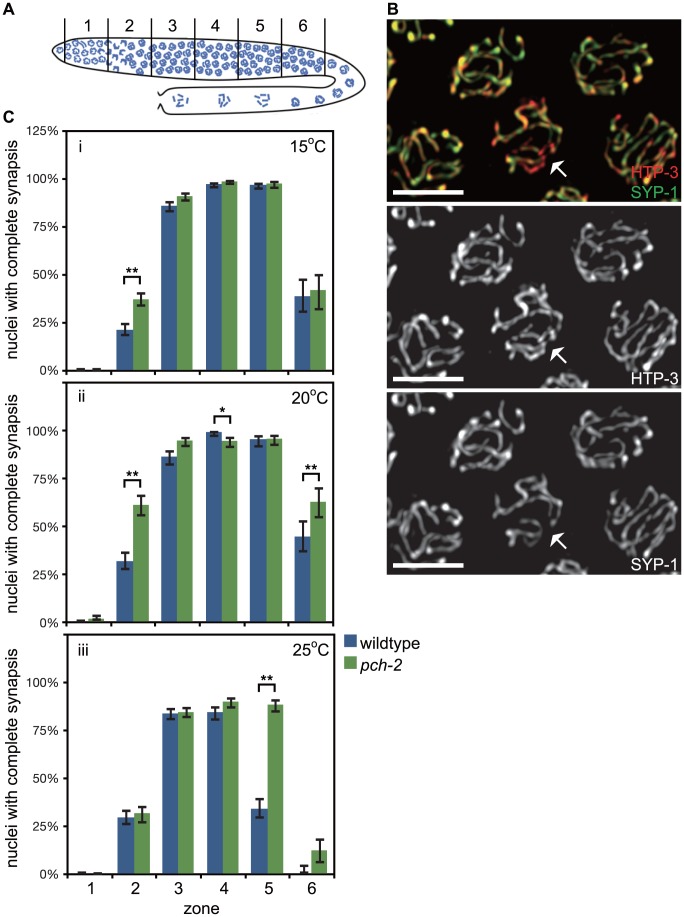
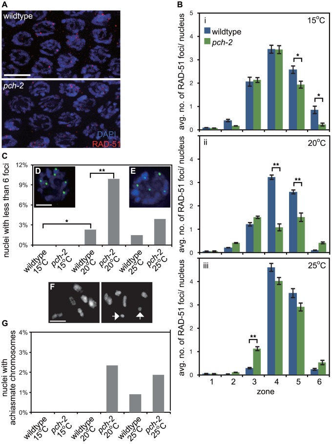
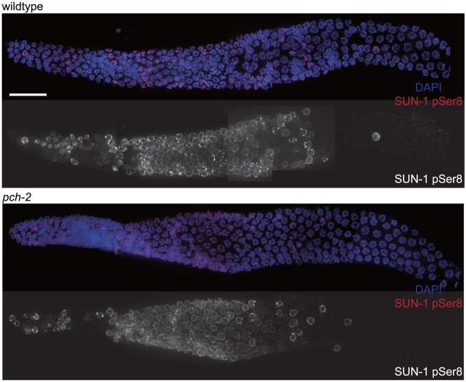
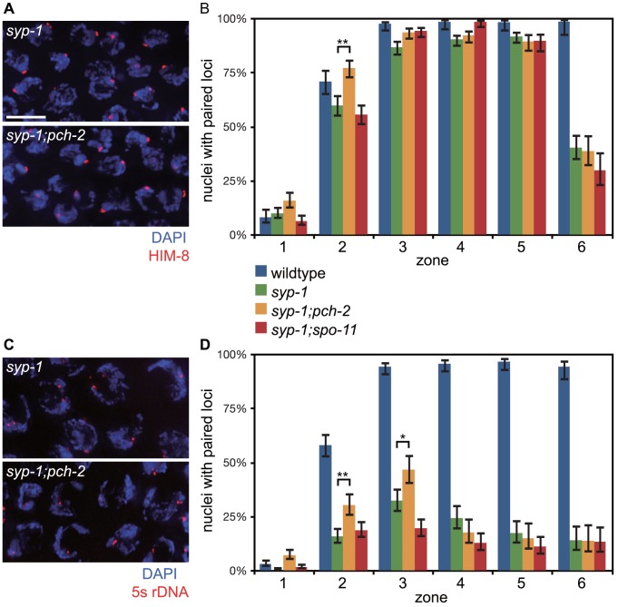
Meiotic chromosome segregation relies on homologous chromosomes being linked by at least one crossover, the obligate crossover. Homolog pairing, synapsis and meiosis specific DNA repair mechanisms are required for crossovers but how they are coordinated to promote the obligate crossover is not well understood. PCH-2 is a highly conserved meiotic AAA+-ATPase that has been assigned a variety of functions; whether these functions reflect its conserved role has been difficult to determine. We show that PCH-2 restrains pairing, synapsis and recombination in C. elegans. Loss of pch-2 results in the acceleration of synapsis and homolog-dependent meiotic DNA repair, producing a subtle increase in meiotic defects, and suppresses pairing, synapsis and recombination defects in some mutant backgrounds. Some defects in pch-2 mutants can be suppressed by incubation at lower temperature and these defects increase in frequency in wildtype worms grown at higher temperature, suggesting that PCH-2 introduces a kinetic barrier to the formation of intermediates that support pairing, synapsis or crossover recombination. We hypothesize that this kinetic barrier contributes to quality control during meiotic prophase. Consistent with this possibility, defects in pch-2 mutants become more severe when another quality control mechanism, germline apoptosis, is abrogated or meiotic DNA repair is mildly disrupted. PCH-2 is expressed in germline nuclei immediately preceding the onset of stable homolog pairing and synapsis. Once chromosomes are synapsed, PCH-2 localizes to the SC and is removed in late pachytene, prior to SC disassembly, correlating with when homolog-dependent DNA repair mechanisms predominate in the germline. Indeed, loss of pch-2 results in premature loss of homolog access. Altogether, our data indicate that PCH-2 coordinates pairing, synapsis and recombination to promote crossover assurance. Specifically, we propose that the conserved function of PCH-2 is to destabilize pairing and/or recombination intermediates to slow their progression and ensure their fidelity during meiotic prophase.


Conflict of interest statement

The authors have declared that no competing interests exist.

Figures

Figure 1
Figure 1. PCH-2's role in regulating synapsis is temperature sensitive.
A. Cartoon of the C. elegans germline divided into six zones of equal size. In all graphs, meiotic progression is from left to right. B. Meiotic nuclei in wildtype worms stained with antibodies against the axial element component HTP-3 (red) and the central element component SYP-1 (green). Most nuclei exhibit complete colocalization on HTP-3 and SYP-1. One nucleus (indicated by arrow) includes stretches of HTP-3 without SYP-1, indicating the presence of unsynapsed chromosomes. Grayscale images of HTP-3 and SYP-1 are also provided. Unless stated otherwise, scale bars in all images represent 5 microns. C. Histograms representing the percentage of nuclei that exhibit complete synapsis as a function of meiotic progression in wildtype and pch-2 mutant worms at 15°C (i), 20°C (ii) and 25°C (iii). Error bars indicate 95% confidence interval. For all graphs, a * indicates a p value<0.05 and a ** indicates a p value of <0.0001. Significance was assessed by performing Fisher's exact test.
Figure 2
Figure 2. PCH-2's role in regulating recombination is temperature sensitive.
A. Meiotic nuclei in early pachytene in wildtype and pch-2 mutant worms stained with DNA (blue) and antibodies against RAD-51 (red). B. Histograms representing the average number of RAD-51 foci per nucleus as a function of meiotic progression in wildtype and pch-2 mutant worms at 15°C (i), 20°C (ii) and 25°C (iii). Error bars indicate standard error of the mean. C. Histogram representing the percentage of nuclei with less than six GFP::COSA-1 foci per nucleus in wildtype and pch-2 mutant worms grown at 15°C, 20°C and 25°C. The number of nuclei assayed for each genotype are as follows: wildtype at 15°C, 303, at 20°C, 304, and 25°C, 339; pch-2 at 15°C, 318, at 20°C, 293, and 25°C, 360. D. A meiotic nucleus stained with DAPI (blue) with six GFP::COSA-1 foci (green). E. A meiotic nucleus stained with DAPI (blue) with five GFP::COSA-1 foci (green). F. Representative images of late meiotic nuclei without (top) and with (bottom) achiasmate chromosomes (arrows). G. Histogram representing the percentage of meiotic nuclei with achiasmate chromosomes in wildtype and pch-2 mutant worms grown at 15°C, 20°C and 25°C. The number of nuclei assayed for each genotype are as follows: wildtype at 15°C, 91, at 20°C, 156, and 25°C, 109; pch-2 at 15°C, 96, at 20°C, 170, and 25°C, 106. For all graphs, a * indicates a p value<0.05 and a ** indicates a p value of <0.0001. Significance was assessed by performing either a paired t-test (2B) or Fisher's exact test (2C).
Figure 3
Figure 3. The appearance and disappearance of phosphorylated SUN-1 is not affected by mutation of pch-2.
Dissected germlines stained with DAPI (blue) and antibodies against SUN-1 pSer8 (red) in wildtype and pch-2 mutant worms. Grayscale images of SUN-1 pSer8 for both genotypes are also provided. Scale bar represents 20 microns.
Figure 4
Figure 4. Mutation of pch-2 suppresses pairing defects in syp-1 mutants.
A. Meiotic nuclei in syp-1 and syp-1;pch-2 mutants stained with DAPI (blue) and antibodies against the X chromosome PC protein HIM-8 (red). B. Histogram representing the percentage of nuclei with paired HIM-8 signals as a function of germline position in wildtype (from Figure S1A), syp-1, syp-1;pch-2, and syp-1;spo-11 mutants. C. Meiotic nuclei in syp-1 and syp-1;pch-2 mutants stained with DAPI (blue) and a FISH probe against the 5 s rDNA locus (red). D. Histogram representing the percentage of nuclei with paired FISH signals as a function of germline position in wildtype (from Figure S1B), syp-1, syp-1;pch-2, and syp-1;spo-11 mutants. Error bars in both graphs indicate 95% confidence intervals. A * indicates a p value<0.05 and a ** indicates a p value of <0.0001. Significance was assessed by performing Fisher's exact test.
Figure 5
Figure 5. Mutation of pch-2 suppresses synapsis defects in meDf2 heterozygotes.
A. Meiotic nuclei in meDf2/+ and meDf2/+;pch-2 double mutants stained with antibodies against HTP-3 (red) and SYP-1 (green). A fraction of meiotic nuclei in meDf2/+ have unsynapsed X chromosomes (arrows). meDf2/+;pch-2 double mutants exhibit complete colocalization of HTP-3 and SYP-1, indicating complete synapsis. Grayscale images of HTP-3 and SYP-1 are also provided for both genotypes. B. Histogram representing the percentage of meiotic nuclei that exhibit complete synapsis as a function of meiotic progression in meDf2/+ and meDf2/+;pch-2. C. Meiotic nuclei in meDf2/+ and meDf2/+;pch-2 stained with a FISH probe against the X chromosome indicates that X chromosomes are paired in meDf2/+;pch-2 mutants. D. Histogram representing the percentage of nuclei with paired X FISH signals as a function of meiotic progression in meDf2/+ and meDf2/+;pch-2 mutant worms. Error bars in both graphs indicate 95% confidence intervals. E. Histogram representing the number of late meiotic nuclei with achiasmate chromosomes in meDf2/+ (54 nuclei) and meDf2/+;pch-2 (78 nuclei). A * indicates a p value<0.05. Significance was assessed by performing Fisher's exact test.
Figure 6
Figure 6. Mutation of pch-2 exacerbates meiotic defects in mutants defective in germline apoptosis and HTP-3 function.
Histogram representing the percentage of late meiotic nuclei in ced-4 (79 nuclei), ced-4;pch-2 (68 nuclei), htp-3 (97 nuclei) and htp-3;pch-2 (107 nuclei) mutants. A * indicates a p value<0.05. Significance was assessed by performing Fisher's exact test.
Figure 7
Figure 7. PCH-2 localizes to the SC when inter-homolog DNA repair mechanisms are active.
A. A wildtype hermaphrodite germline stained with DAPI and antibodies against PCH-2. PCH-2 is present in germline nuclei prior to the transition zone and removed in late pachytene. Scale bar represents 20 microns. B. Meiotic nuclei in the transition zone stained with DAPI (blue) and antibodies against PCH-2 (green). Meiotic progression for both A and B is from left to right. C. Meiotic nuclei in mid-pachytene stained with antibodies against SYP-1 (red) and PCH-2 (green). PCH-2 colocalizes with SYP-1. D. Meiotic nuclei in late pachytene stained with antibodies against SYP-1 (red) and PCH-2 (green). PCH-2 is absent from meiotic chromosomes. E. Meiotic nuclei in syp-1 mutants stained with antibodies against HTP-3 (red) and PCH-2 (green). F. Meiotic nuclei in htp-1 mutants stained with antibodies against HTP-3 (red) and PCH-2 (green). G. Meiotic nuclei in mpk-1 mutants stained with antibodies against SYP-1 (red) and PCH-2 (green). H. Meiotic nuclei in mpk-1 mutants in which dpl-1 has been knocked down by RNAi stained with antibodies against SYP-1 (red) and PCH-2 (green). I. Meiotic nuclei in late pachytene in msh-5 mutants stained with antibodies against SYP-1 (red) and PCH-2 (green). J. Meiotic nuclei in late pachytene in rtel-1 mutants stained with antibodies against HTP-3 (red) and PCH-2 (green).
Figure 8
Figure 8. pch-2 is required to maintain inter-homolog DNA repair in mid-pachytene.
A. Histogram representing the average number of RAD-51 foci per nucleus as a function of meiotic progression in syp-1 and pch-2;syp-1 mutant worms. Error bars indicate standard error of the mean. A ** indicates a p value<0.0001. Significance was assessed by performing a paired t-test. B. Mos1 excision based DSB repair assay. C. Time-course analysis of inter-homolog recombination induced at the Mos1 site. Chart shows the frequency of unc-5(+) recombinants among progeny derived from eggs laid in the indicated time intervals (hours phs) and the frequency of CO and NCO repair types. D. Genetic analysis of meiotic recombination in wildtype and pch-2 mutants. Physical and genetic maps of Chromosome III and the X chromosome are depicted to scale. Genetic distance is shown in centimorgans and was calculated as number of recombinants divided by total number of chromosomes assayed. For both C and D, a * indicates a p value<0.05 and significance was assessed by performing Fischer's exact test.
Figure 9
Figure 9. Mutation of pch-2 reduces the percentage of nuclei with six GFP::COSA-1 foci in meDf2 homozygotes.
A. A dissected germline stained with DAPI (blue) and antibodies against SUN-1 pSer8 (red) and PCH-2 (green). The extension of PCH-2 staining into late pachytene correlates with the extension of phosphorylated SUN-1 staining. Scale bar represents 20 microns. B. Histogram representing the percentage of meiotic nuclei that contain a given number of GFP::COSA-1 foci in meDf2 and meDf2;pch-2 mutants. 174 meDf2 nuclei and 178 meDf2;pch-2 nuclei were counted. Error bars represent 95% confidence interval. C. Histogram representing the average number of RAD-51 foci per nucleus in meDf2 and meDf2;pch-2 mutants. Error bars indicate standard error of the mean. For both graphs, a * indicates a p value<0.05. Significance was assessed by performing either Fisher's exact test (7B) or a paired t-test (7C). D. Genetic analysis of meiotic recombination in wildtype, pch-2, meDf2 and meDf2;pch-2 mutants represented as fractions of wildtype recombination and color-coded according to the legend on the right.
Figure 10
Figure 10. Model for role of PCH-2 during crossover formation.
See discussion for details.

Similar articles

Cited by

References

    1. Baudat F, de Massy B (2007) Regulating double-stranded DNA break repair towards crossover or non-crossover during mammalian meiosis. Chromosome Res 15: 565–577. - PubMed
    1. Martinez-Perez E, Colaiacovo MP (2009) Distribution of meiotic recombination events: talking to your neighbors. Curr Opin Genet Dev 19: 105–112. - PMC - PubMed
    1. Bhalla N, Dernburg AF (2008) Prelude to a division. Annu Rev Cell Dev Biol 24: 397–424. - PMC - PubMed
    1. Borner GV, Kleckner N, Hunter N (2004) Crossover/noncrossover differentiation, synaptonemal complex formation, and regulatory surveillance at the leptotene/zygotene transition of meiosis. Cell 117: 29–45. - PubMed
    1. Bishop DK, Zickler D (2004) Early decision; meiotic crossover interference prior to stable strand exchange and synapsis. Cell 117: 9–15. - PubMed

Publication types

Substances