. 2014 Jun;124(6):2441-55.
doi: 10.1172/JCI70522. Epub 2014 Apr 24.

Intrinsic TGF-β signaling promotes age-dependent CD8+ T cell polyfunctionality attrition

Intrinsic TGF-β signaling promotes age-dependent CD8+ T cell polyfunctionality attrition

Rajarshi Bhadra et al. J Clin Invest. 2014 Jun.

Abstract

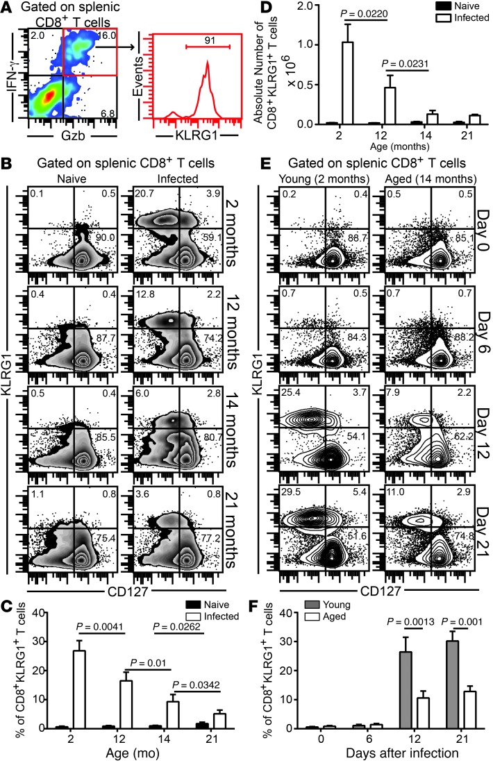
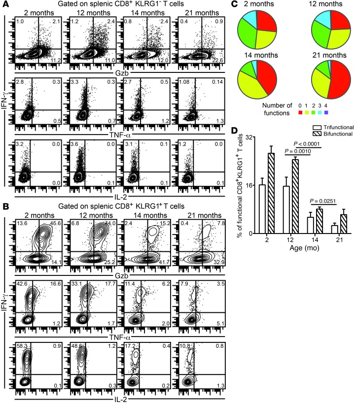
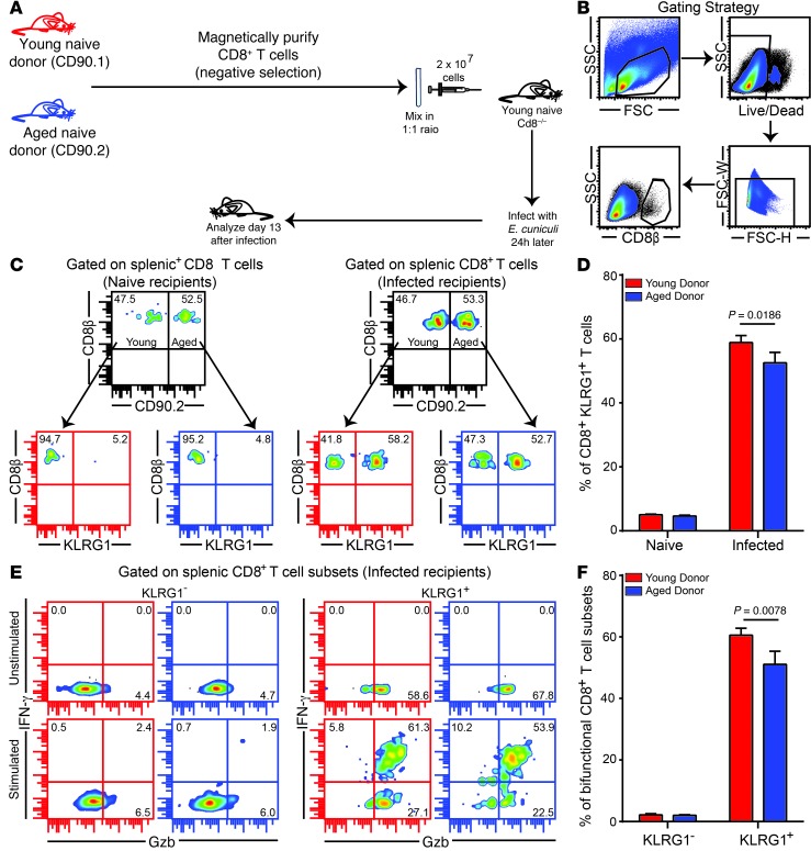
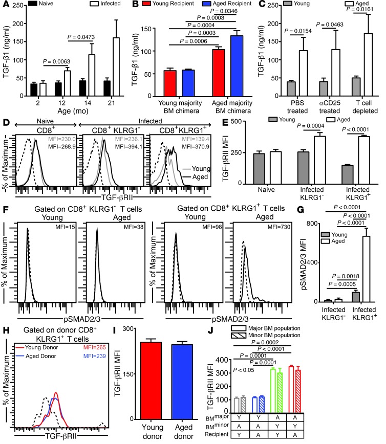
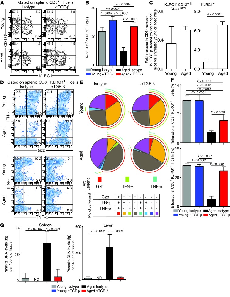
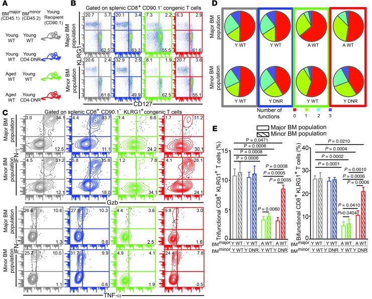
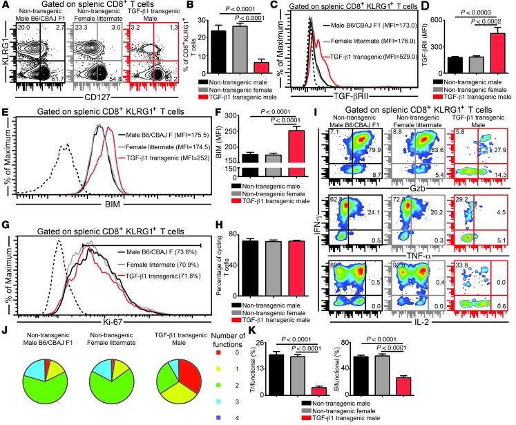
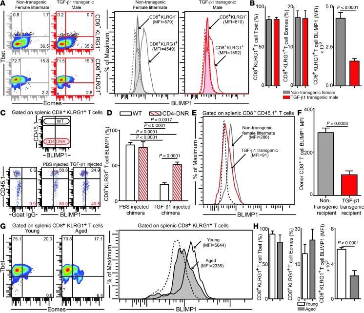
Advanced age is associated with immune system deficits that result in an increased susceptibility to infectious diseases; however, specific mediators of age-dependent immune dysfunction have not been fully elucidated. Here we demonstrated that aged mice exhibit poor effector CD8+ T cell polyfunctionality, primarily due to CD8+ T cell-extrinsic deficits, and that reduced CD8+ T cell polyfunctionality correlates with increased susceptibility to pathogenic diseases. In aged animals challenged with the parasite Encephalitozoon cuniculi, effector CD8+ T cell survival and polyfunctionality were suppressed by highly elevated TGF-β1. Furthermore, TGF-β depletion reduced effector CD8+ T cell apoptosis in both young and aged mice and enhanced effector CD8+ T cell polyfunctionality in aged mice. Surprisingly, intrinsic blockade of TGF-β signaling in CD8+ T cells was sufficient to rescue polyfunctionality in aged animals. Together, these data demonstrate that low levels of TGF-β1 promote apoptosis of CD8+ effector T cells and high TGF-β1 levels associated with age result in both CD8+ T cell apoptosis and an altered transcriptional profile, which correlates with loss of polyfunctionality. Furthermore, elevated TGF-β levels are observed in the elderly human population and in aged Drosophila, suggesting that TGF-β represents an evolutionarily conserved negative regulator of the immune response in aging organisms.


Figures

Figure 1
Figure 1. Attrition of effector CD8+KLRG1+ T cells in E. cuniculi–challenged aged mice.
(A) Young mice were orally infected with E. cuniculi spores, and KLRG1 expression was assessed at day 12 after infection in IFN-γ+Gzb+ splenic CD8+ T cells. (BD) Frequency (B and C) and absolute number (D) of CD8+KLRG1+ T cells in E. cuniculi–challenged animals at 2, 12, 14, and 21 months of age. (E and F) KLRG1+ CD8+ kinetic was assessed in young and 14-month-old mice at days 0, 6, 12, and 21 after infection. Data represent 2 experiments with at least 4 mice per group. Numbers in dot plots and histograms denote percentages.
Figure 2
Figure 2. Downregulation of effector CD8+ T cell polyfunctionality in E. cuniculi–challenged aged mice.
(A and B) KLRG1 (A) and KLRG1+ (B) effector CD8+ T cells from mice at various ages were assayed for IFN-γ, Gzb, TNF-α, and IL-2 production at day 12 after infection. (C) Proportion of polyfunctional (IFN-γ, Gzb, TNF-α, and/or IL-2) KLRG1+ CD8+ T cells in these mice. (D) Percentage of KLRG1+ CD8+ T cells exhibiting 2 or 3 functions (IFN-γ, Gzb, and/or TNF-α), shown as bar graphs. Data represent 2 experiments with at least 4 mice per group. Numbers in dot plots denote percentage.
Figure 3
Figure 3. Poor effector CD8+KLRG1+ T cell functionality is not primarily caused by CD8+ T cell–intrinsic deficits.
(A) Equal number of CD8+ T cells from CD90.1 young (6–8 weeks old) and CD90.2 aged (14 months old) naive mice were adoptively transferred to young Cd8–/– mice. 24 hours later, recipients were challenged with E. cuniculi, and CD8+ T cell response was assessed after 12 days of infection. (B) Gating strategy. (C and D) Donor CD8+KLRG1+ T cell frequencies in recipient mice. (E and F) IFN-γ and Gzb production were evaluated in donor CD8+KLRG1+ and CD8+KLRG1 T cells in these animals. Data represent 2 experiments with 3–4 mice per group. Unless otherwise indicated, “aged” refers to 14- to 15-month-old mice throughout. Numbers in dot plots denote percentages.
Figure 4
Figure 4. Plasma TGF-β1 is highly elevated in E. cuniculi–challenged aged mice.
(A) TGF-β1 plasma levels in parasite-infected mice of various ages at day 12 after infection. (B) TGF-β plasma levels in E. cuniculi–challenged young majority and aged majority BM chimeras generated in young or aged recipients. (C) Plasma collected from young (6–8 weeks old) or aged (14–15 months old) mice treated with PBS, with anti-CD25, or with anti-thymocyte to deplete T cells was assayed for TGF-β. (D and E) TGF-βRII expression was measured on splenic CD8+ subsets in young (6–8 weeks old) and aged (14–15 months old) mice by polychromatic flow cytometry. (F and G) Direct ex vivo SMAD2/3 phosphorylation on splenic CD8+ subsets. (H and I) Young (CD90.1; 6–8 weeks old) and aged (CD90.2; 14 months old) CD8+ T cells from naive donors were adoptively transferred into young Cd8–/– recipients, followed by parasite challenge. TGF-βRII expression was evaluated in the recipients on donor effector CD8+KLRG1+ T cells in spleen. (J) TGF-βRII expression levels on splenic effector CD8+KLRG1+ T cells in young majority or aged majority BM chimeras formed in young or aged recipients. Y, young; A, aged. Data represent 2 experiments with 4 mice per group. Numbers in histograms denote MFI.
Figure 5
Figure 5. Anti–TGF-β treatment rescues effector CD8+ T cell polyfunctionality in E. cuniculi–infected aged mice.
(A and B) KLRG1 expression on splenic CD8+ T cells was evaluated in young (6–8 weeks old) or aged (14–15 months old) mice treated with isotype control antibody or anti–TGF-β at day 12 after infection. (C) Fold increase in absolute numbers of CD8+ T cell subsets in anti–TGF-β–treated young or aged mice compared with the respective isotype-treated controls. (DF) IFN-γ, Gzb, and TNF-α production by effector CD8+KLRG1+ T cells was evaluated by 6-color flow cytometry. (G) Parasite DNA levels in spleen and liver was assessed in these mice by quantitative PCR. Data are representative of 3 experiments (AF) or 2 independent experiments (G) with 4–6 mice per group. Numbers in dot plots denote percentages.
Figure 6
Figure 6. CD8+ T cell–intrinsic TGF-β signaling results in poor development of E. cuniculi–specific effector CD8+ T cell response in aged mice.
(A) Mixed bone marrow chimera generation using a combination of young or aged WT and young CD4-DNR donors. (B) Percentage of splenic KLRG1+ CD8+ T cells was evaluated in these chimeras at day 12 after infection. (CE) Effector CD8+ T cell polyfunctionality (IFN-γ, Gzb, and/or TNF-α) was assayed in chimeric mice by polychromatic flow cytometry. Data represent 2 experiments with 3–4 chimeras per group. Numbers in dot plots denote percentages.
Figure 7
Figure 7. Elevated TGF-β levels in young TGF-β transgenic mice downregulates effector CD8+ T cell polyfunctionality.
(A and B) KLRG1 expression was assessed on splenic CD8+ T cells in E. cuniculi–infected young Alb–TGF-β1 mice and control groups (parental and littermate) at day 12 after infection. (C and D) TGF-βRII expression on effector CD8+KLRG1+ T cells, presented as histograms and bar graphs. (E and F) Intracellular BIM level in the KLRG1+ subset, assessed by flow cytometry. (G and H) Frequency of proliferating effector CD8+ T cells. (I and J) IFN-γ, Gzb, TNF-α, and IL-2 production by effector CD8+KLRG1+ T cells, assayed by 7-color flow cytometry. (K) Percentage of KLRG1+ CD8+ T cells showing 2 or more functions (IFN-γ, Gzb, and/or TNF-α), shown as bar graphs. Data represent 3 independent experiments with 4 mice per group. Numbers in histograms denote MFI (C and E) or percentage (G); numbers in dot plots denote percentages.
Figure 8
Figure 8. Highly elevated TGF-β levels downregulate BLIMP1 expression on effector CD8+KLRG1+ T cells in the E. cuniculi model.
(A and B) Expression levels of transcription factors Tbet, Eomes, and BLIMP1 in splenic CD8+KLRG1+ and CD8+KLRG1 T cells in young TGF-β1 transgenic mice and littermate controls. (C and D) Acutely infected Young WT:Young CD4-DNR BM chimeras were treated i.v. with 500 ng murine TGF-β1 or PBS. BLIMP1 level was then evaluated in KLRG1+ effectors at day 12 after infection. (E and F) YFP+CD8+ effectors were sorted from E. cuniculi–challenged IFN-γ reporter mice (CD45.1; day 12 after infection) and adoptively transferred to Alb–TGF-β1 recipients or littermate controls at day 7 after infection. BLIMP1 expression in donor cells was assessed 5 days after transfer. (G and H) BLIMP1 level in KLRG1+ CD8+ T cells in young or aged mice at day 12 after infection. Data represent at least 2 experiments with 4–5 mice per group. Numbers in dot plots represent percentages; numbers in histograms represent MFI.

References

    1. Nikolich-Zugich J. The aging immune system: Challenges for the 21st century. Semin Immunol. 2012;24(5):301–302. doi: 10.1016/j.smim.2012.09.001. - DOI - PMC - PubMed
    1. Nikolich-Zugich J. T cell aging: naive but not young. J Exp Med. 2005;201(6):837–840. doi: 10.1084/jem.20050341. - DOI - PMC - PubMed
    1. Yager EJ, Ahmed M, Lanzer K, Randall TD, Woodland DL, Blackman MA. Age-associated decline in T cell repertoire diversity leads to holes in the repertoire and impaired immunity to influenza virus. J Exp Med. 2008;205(3):711–723. doi: 10.1084/jem.20071140. - DOI - PMC - PubMed
    1. Seder RA, Darrah PA, Roederer M. T-cell quality in memory and protection: implications for vaccine design. Nat Rev Immunol. 2008;8(4):247–258. doi: 10.1038/nri2274. - DOI - PubMed
    1. Precopio ML, et al. Immunization with vaccinia virus induces polyfunctional and phenotypically distinctive CD8(+) T cell responses. J Exp Med. 2007;204(6):1405–1416. doi: 10.1084/jem.20062363. - DOI - PMC - PubMed

Publication types

MeSH terms