. 2014 Jun;124(6):2560-70.
doi: 10.1172/JCI72063. Epub 2014 Apr 24.

Peripheral nervous system plasmalogens regulate Schwann cell differentiation and myelination

Peripheral nervous system plasmalogens regulate Schwann cell differentiation and myelination

Tiago Ferreira da Silva et al. J Clin Invest. 2014 Jun.

Abstract

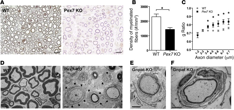
Rhizomelic chondrodysplasia punctata (RCDP) is a developmental disorder characterized by hypotonia, cataracts, abnormal ossification, impaired motor development, and intellectual disability. The underlying etiology of RCDP is a deficiency in the biosynthesis of ether phospholipids, of which plasmalogens are the most abundant form in nervous tissue and myelin; however, the role of plasmalogens in the peripheral nervous system is poorly defined. Here, we used mouse models of RCDP and analyzed the consequence of plasmalogen deficiency in peripheral nerves. We determined that plasmalogens are crucial for Schwann cell development and differentiation and that plasmalogen defects impaired radial sorting, myelination, and myelin structure. Plasmalogen insufficiency resulted in defective protein kinase B (AKT) phosphorylation and subsequent signaling, causing overt activation of glycogen synthase kinase 3β (GSK3β) in nerves of mutant mice. Treatment with GSK3β inhibitors, lithium, or 4-benzyl-2-methyl-1,2,4-thiadiazolidine-3,5-dione (TDZD-8) restored Schwann cell defects, effectively bypassing plasmalogen deficiency. Our results demonstrate the requirement of plasmalogens for the correct and timely differentiation of Schwann cells and for the process of myelination. In addition, these studies identify a mechanism by which the lack of a membrane phospholipid causes neuropathology, implicating plasmalogens as regulators of membrane and cell signaling.


Figures

Figure 1
Figure 1. Plasmalogen deficiency causes defects in axonal sorting and myelination.
(A) Electron microscopic analysis of sciatic nerves from P5 and P15 WT and Gnpat-KO mice. Bundles in P5 Gnpat-KO nerves contained large axons (asterisks), whereas in WT nerves these axons had been sorted (arrowhead), and the bundles contained very small-caliber axons (arrow). At P15, sciatic nerves from Gnpat-KO mice had Remak bundles with only 1 axon (arrowheads). Scale bars: 2 μm. (B) Composition of axon bundles in sciatic nerves from P5 WT, Pex7-KO, and Gnpat-KO mice. *P = 0.031; **P = 0.011. (C) Density of sorted axons in sciatic nerves from P5 WT, Pex7-KO, and Gnpat-KO mice. *P = 0.003. (D) Composition of Remak bundles in nerves from adult WT and Pex7-KO mice. *P = 0.013. (E) Density of unmyelinated fibers (UMF) in Remak bundles of nerves from adult WT and Pex7-KO mice. (F) Quantification of myelin thickness by g ratio in sciatic nerves at P15. Results are graphed as boxes with a line at the mean and whiskers from the minimal to maximal values. *P = 0.005. (G) DRG cocultures of neurons and Schwann cells from WT and Gnpat-KO mice stained for neuronal βII-tubulin (green) and for the myelin protein MBP (red). Scale bars: 200 μm. (H) Density of myelin segments in DRG cocultures from WT and Gnpat-KO mice. *P = 0.001. (I) Length of individual myelin segments in myelinating cocultures. *P = 0.001.
Figure 2
Figure 2. Plasmalogens and MBP coordinate myelination.
(A) Quantification of myelin thickness by g ratio in sciatic nerves from 3-month-old WT and Pex7-KO mice. Results are the mean ± SEM. (B) X-ray diffraction analysis of sciatic nerves from WT and Pex7-KO mice. Results are graphed as the mean ± SEM. P = 0.01. (C) Immunofluorescence analysis of teased fibers from adult WT and Gnpat-KO nerves stained with F-actin (red). Arrows point to Schmidt-Lanterman incisures. Scale bars: 10 μm. (D) Immunofluorescence analysis of sciatic nerve teased fibers stained with an antibody against DRP2 (green) showing abnormal apposition in mutant nerves. Scale bars: 10 μm. (E) PPD-stained cross sections of sciatic nerves from P30 mice. Scale bars: 25 μm. (F) Quantification of the degree of myelination by g ratio in sciatic nerves from P30 mice. Results are graphed as boxes with a line at the mean and whiskers from the minimal to maximal values. *P < 0.001. (G) Calculated motor nerve conduction velocities (MNCV) in 3-month-old WT, shi, Pex7-KO, Pex7:shi DM, Gnpat-KO, and Gnpat:shi DM mice. *P < 0.0001; #P = 0.001.
Figure 3
Figure 3. Plasmalogens are important players during remyelination of the PNS.
(A) PPD-stained cross sections of the distal segment of sciatic nerves 15 days after nerve crush. Scale bars: 10 μm. (B) Degree of regeneration as measured by the density of myelinated axons in the distal segment 15 days after sciatic nerve crush. *P = 0.014. (C) Extent of impaired regeneration as measured by g ratio determination. Results are graphed as boxes with a line at the mean and whiskers from the minimal to maximal values. *P = 0.029. (D) Electron microscopic analysis of the distal segment of crushed sciatic nerves from WT and Pex7-KO mice. In WT nerves, remyelination of regenerating axons was evident (arrowheads), whereas Pex7-KO axons of a similar caliber were devoid of myelin (asterisks). Scale bars: 5 μm. (E) Density of axons lacking myelin (demyelinated axons) in the distal segment following sciatic nerve crush. Error bars represent SEM. *P = 0.012.
Figure 4
Figure 4. Deficiency in plasmalogens impairs the ability of Schwann cells to sustain myelinated axons.
(A) PPD-stained cross sections of sciatic nerves from 1.5-year-old WT and Pex7-KO mice revealing demyelination and loss of axons in mutant nerves. Scale bars: 10 μm. (B) Density of myelinated axons in sciatic nerves from WT and Pex7-KO mice. Error bars represent SEM. *P = 0.012. (C) Quantification of the degree of myelination by g ratio in sciatic nerves from 1.5-year-old WT and Pex7-KO mice. *P = 0.026. Error bars represent SEM. (D) Electron microscopic analysis of sciatic nerves from representative 1.5-year-old WT and Pex7-KO mice showing an axon devoid of myelin (asterisk), severely demyelinated axons (arrows), and the presence of extended Schwann cell processes (arrowheads) throughout the perineurium. Scale bars: 2 μm. (E) Schwann cell from an aged Gnpat-KO mouse undergoing remyelination of a demyelinated axon. Scale bar: 2 μm. (F) Engulfment of a demyelinated axon by 2 Schwann cells, generating concentric deposition of membrane processes, similar to the formation of onion bulbs. Scale bar: 2 μm.
Figure 5
Figure 5. Defects in plasmalogens result in impaired AKT activation and signaling.
(A) Western blot analysis and quantification of AKT phosphorylation (p-AKT) in sciatic nerve lysates of P15 WT and Gnpat-KO mice. *P = 0.018; **P = 0.006. (BE) Quantification of phosphorylated forms of GSK3β at Ser9 (B), c-RAF at Ser259 (C), PDK1 at Ser241 (D), and PTEN at Ser380 (E) in sciatic nerves from WT and Gnpat-KO mice. *P < 0.02. (F) Density of BrdU-positive cells in nerves from P4 WT and Gnpat-KO mice. *P = 0.020. (G) Western blot analyses of p-AKT and p-ERK1/2 in serum-starved MEFs from WT and Gnpat-KO mice stimulated with 10% FBS. (H and I) Quantification of p-AKT at Ser473 (H) and Thr308 (I) in primary WT and Gnpat-KO Schwann cells after stimulation with NRG1. (J) Western blot analysis of total and p-AKT in cytosolic and membrane fractions of serum-starved MEFs from WT and Gnpat-KO mice stimulated with 10% FBS. Western blot analysis of caveolin 1 (CAV1), GAPDH, and peroxisomal thiolase (ACAA1) used to control membrane fractions and cytosolic fractions and to monitor lack of solubilized organelles in cytosolic fractions, respectively. (K) DRG cocultures from WT and Gnpat-KO mice treated with DMSO (control) or with SC79, stained for neuronal βIII-tubulin (green) and MBP (red). Scale bars: 100 μm. (L) Density of myelin segments in DRG cocultures from WT and Gnpat-KO mice after DMSO and SC79 treatment. *P < 0.002. (M) Length of individual myelin segments in myelinating cocultures. *P < 0.01. Error bars represent SEM in all graphs.
Figure 6
Figure 6. Treatment with GSK3β inhibitors restores Schwann cell differentiation and radial sorting defects.
(A) In vitro myelination after treatment with NaCl (control) or LiCl. *P = 0.001. (B) Strategy for in vivo treatments. For the assessment of LiCl on the phosphorylation of AKT and GSK3β, mice were injected with LiCl on alternating days from P7 to P15 (upper diagram). For the assessment of LiCl on nerve pathology, mice were injected daily with LiCl from P1 to P6 (lower diagram). (C and D) Quantification of p-GSK3β at Tyr216 (*P = 0.04) (C) and Ser9 (*P = 0.017) (D) in nerves from P15 mice. (E and F) Quantification of p-AKT at Ser473 (E) and Thr308 (F) in nerves from P15 mice. *P = 0.01. (G and H) Quantification of SOX2 (G) and OCT6 (H) levels in nerves from P6 WT and Gnpat-KO mice. *P = 0.03 (G); P = 0.038 (H). (I) Ultrastructural analysis of sciatic nerves from P6 Gnpat-KO mice treated with NaCl and LiCl. Lines and asterisks indicate axon bundles on control and lithium-treated nerves, respectively. Scale bars: 5 μm. (J) Number of axons in bundles of sciatic nerves from P6 mice treated with NaCl and LiCl. *P = 0.0013. (K) Density of sorted axons (promyelinating stage) in sciatic nerves from P6 mice treated with NaCl and LiCl. *P = 0.0022. (L) Number of axons in bundles of sciatic nerves from P4 mice treated with DMSO and TDZD-8. *P < 0.007. (M) Density of sorted axons in sciatic nerves from P4 WT and Gnpat-KO mice treated with DMSO and TDZD-8. *P < 0.03.
Figure 7
Figure 7. Schematic representation of a proposed model depicting the consequences of plasmalogen deficiency in the PNS.
The deficiency in plasmalogens at the plasma membrane of Schwann cells causes reduced phosphorylation of AKT, leading to an overt activation of GSK3β by reducing the inhibitory phosphorylation at Ser9. Active GSK3β inhibits Schwann cell differentiation, impairing axonal sorting and myelination. Lithium administration to inhibit GSK3β is able to rescue Schwann cell differentiation and maturation in the absence of plasmalogens. During aging, lack of plasmalogens causes demyelination and axonal loss, and Schwann cells extend processes in failed attempts to remyelinate existing axons. Plasmalogens are depicted by gray-colored phospholipids; ҂, impaired signaling; →, induction; ┬, inhibition.

References

    1. Lessig J, Fuchs B. Plasmalogens in biological systems: their role in oxidative processes in biological membranes, their contribution to pathological processes and aging and plasmalogen analysis. Curr Med Chem. 2009;16(16):2021–2041. doi: 10.2174/092986709788682164. - DOI - PubMed
    1. Wanders RJ, Waterham HR. Biochemistry of mammalian peroxisomes revisited. Annu Rev Biochem. 2006;75:295–332. doi: 10.1146/annurev.biochem.74.082803.133329. - DOI - PubMed
    1. Ofman R, Hettema EH, Hogenhout EM, Caruso U, Muijsers AO, Wanders RJ. Acyl-CoA:dihydroxyacetonephosphate acyltransferase: cloning of the human cDNA and resolution of the molecular basis in rhizomelic chondrodysplasia punctata type 2. Hum Mol Genet. 1998;7(5):847–853. doi: 10.1093/hmg/7.5.847. - DOI - PubMed
    1. de Vet EC, Ijlst L, Oostheim W, Wanders RJ, van den Bosch H. Alkyl-dihydroxyacetonephosphate synthase. Fate in peroxisome biogenesis disorders and identification of the point mutation underlying a single enzyme deficiency. J Biol Chem. 1998;273(17):10296–10301. doi: 10.1074/jbc.273.17.10296. - DOI - PubMed
    1. Motley AM, et al. Mutational spectrum in the PEX7 gene and functional analysis of mutant alleles in 78 patients with rhizomelic chondrodysplasia punctata type 1. Am J Hum Genet. 2002;70(3):612–624. doi: 10.1086/338998. - DOI - PMC - PubMed

Publication types

MeSH terms

Substances

Supplementary concepts