. 2014;13(12):1948-57.
doi: 10.4161/cc.28898. Epub 2014 Apr 24.

S-nitrosylation of FLICE inhibitory protein determines its interaction with RIP1 and activation of NF-κB

Affiliations

S-nitrosylation of FLICE inhibitory protein determines its interaction with RIP1 and activation of NF-κB

Siera Jo Talbott et al. Cell Cycle. 2014.

Abstract

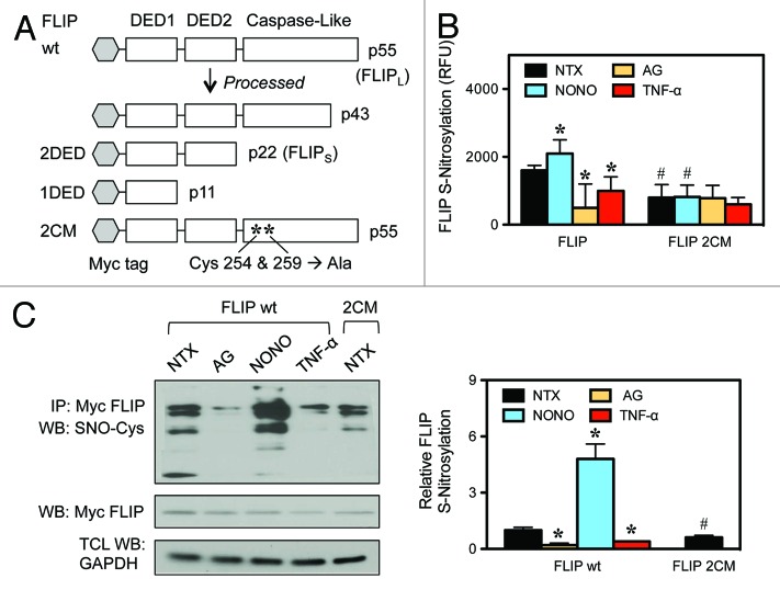
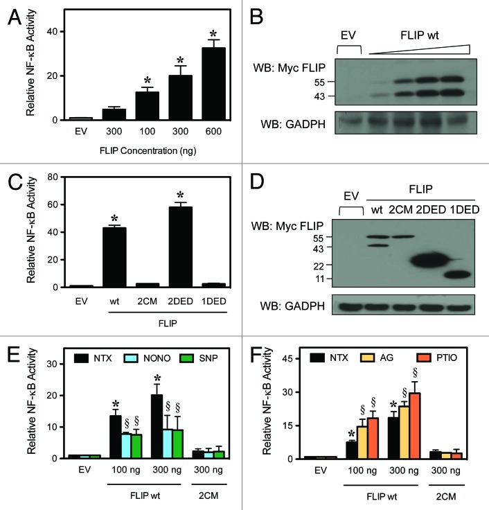
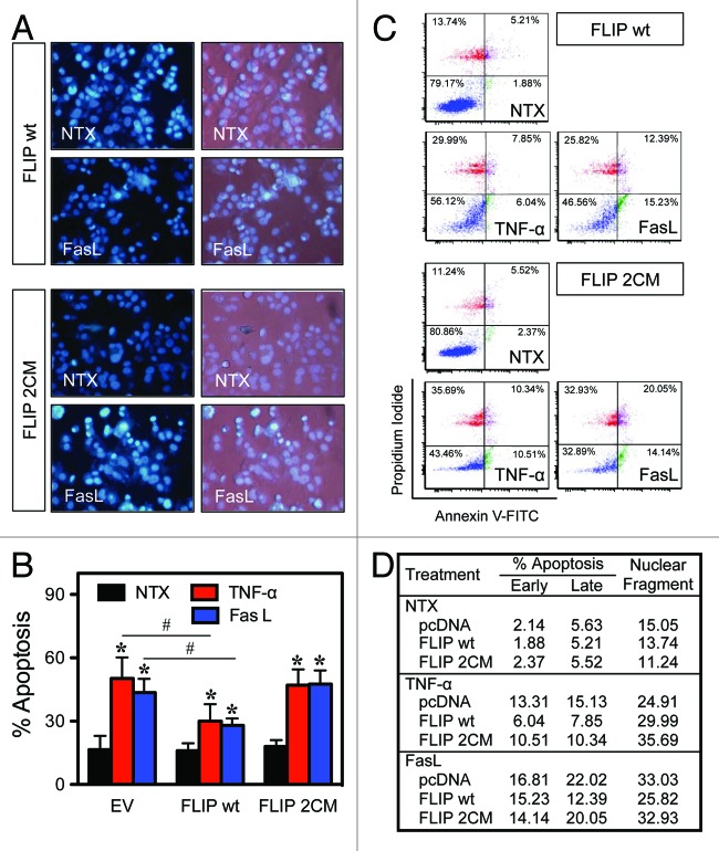
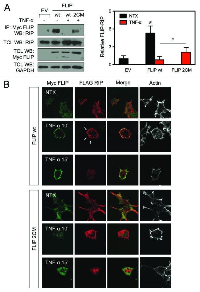
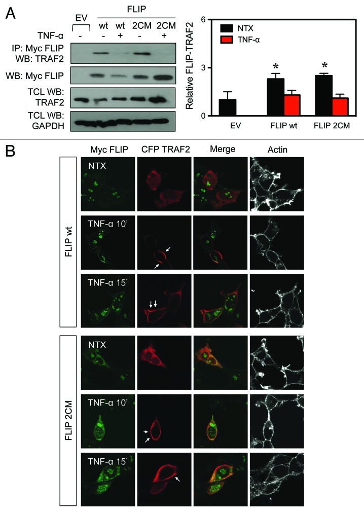
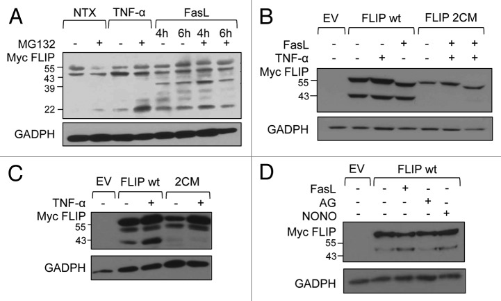
Death receptor (DR) ligation can lead to divergent signaling pathways causing either caspase-mediated cell death or cell proliferation and inflammation. These variations in cellular fate are determined by adaptor proteins that are recruited to the DR signaling complex. FLICE inhibitory protein (FLIP) is an established inhibitor of caspase-8-mediated apoptosis, and it is also involved in NF-κB activation. However, the molecular mechanism that regulates FLIP within this complex is unknown. In this study, we provide new evidence for the regulation of NF-κB by FLIP through S-nitrosylation, which involves covalent modification of the protein's cysteine thiol by nitric oxide to form S-nitrosothiol. Point mutations of FLIP at cysteine residues 254 and 259 prevent FLIP S-nitrosylation and its ability to activate NF-κB. The mechanism by which FLIP nitrosylation regulates NF-κB activity involves RIP1 binding and redistribution, whereas TRAF2 binding and distribution are unaffected. We further show that FLIP processing and cleavage is dependent on its nitrosylation status. Collectively, our study reveals a novel pathway for FLIP regulation of NF-κB through protein S-nitrosylation, which is a key posttranslational mechanism controlling DR-mediated cell death and survival. Since increased expression of FLIP and nitric oxide are frequently observed in chemotherapy-resistant tumors, S-nitrosylation of FLIP could be a key mechanism of chemoresistance and tumor growth.

Keywords: FLIP; NF-κB; RIP1; S-nitrosylation; cancer.


Figures

None
Figure 1. S-nitrosylation of FLIP. (A) A schematic of the domain structure of FLIP isoforms and expression vectors is shown. FLIP wild-type (wt), FLIP death effector domains (2DED), FLIP double cysteine mutant (2CM). (B) HEK-293 cells were transiently transfected with myc-FLIP wt or myc-FLIP 2CM and treated with media alone (NTX), 300 μM AG, 400 μM NONOate for 12 h or 50 ng/mL TNF-α for 30 min. Analysis for S-nitrosylation was determined by fluorescence described in “Materials and Methods”. (C) HEK-293 cells were transiently transfected with myc-FLIP wt or myc-FLIP 2CM. myc-FLIP-expressing cells were treated as in (B). Lysates were coimmunoprecipitated using anti-myc antibodies and assayed for S-nitrocysteine (SNO-Cys) by streptavidin using an S-nitrosylation kit. Membranes were stripped and reprobed with anti-myc antibodies to detect FLIP. Total cell lysates (TCL) were also probed with anti-GAPDH antibodies to control for loading. Data are mean ± SD (n = 3). *P < 0.05 vs. non-treated (NTX) FLIP wt transfected cells. #P < 0.05 vs. FLIP wt transfected cells.
None
Figure 2. FLIP-mediated NF-κB activation. HEK-293 cells were transiently transfected with an NF-κB-luciferase and Renilla-luciferase reporter and expression constructs for the FLIP domains, as indicated. Luciferase activity was determined by the dual-luciferase reporter assay. (A) Cells were transiently transfected with increasing amounts of FLIP wt and an NF-κB luciferase reporter plasmid. NF-κB activity was determined by luminescence according to “Materials and Methods”. (B) Total cell lysates were also analyzed by western blot using an anti-myc antibody to verify the increasing expression levels of FLIP. (C) Cells were transiently transfected with empty vector (EV), FLIP wt, FLIP 2CM, FLIP 2DED, or FLIP 1DED and analyzed for NF-κB activity as noted in (A). (D) Total cell lysates were also analyzed by western blotting as above to verify expression of all constructs. (E) Cells were transiently transfected with EV, FLIP wt or FLIP 2CM. Cells treated with media alone, 400 μM NONOate, or 500 μM SNP for 12 h to supplement nitric oxide and analyzed for NF-κB activity as noted in (A). (F) Cells were transiently transfected with EV, FLIP wt, or FLIP 2CM, and cells were treated with media alone, 300 μM AG or 300 μM PTIO for 12 h to inhibit NO production. Lysates were analyzed for NF-κB activity as noted in (A). Data are mean ± SD (n = 3). *P < 0.05 vs. non-treated (NTX) EV-transfected cells. §P < 0.05 vs. non-treated (NTX) control.
None
Figure 3. S-nitrosylation of FLIP modulates its anti-apoptotic activity. MCF-7 cells were transiently transfected with FLIP wt or FLIP 2CM expression constructs. Twenty-four hours post-transfection, cells were treated with media alone, 50 ng/mL TNF-α, or 200 ng/mL FasL for 16 h, and apoptosis was determined by Hoechst assay. (A) Representative fluorescence micrographs of treated cells stained with the Hoechst dye. (B) Quantitative analysis of 15 fields of view. (C) Cells were similarly treated with TNF-α and FasL for 16 h, and apoptosis was determined by flow cytometry using annexin V and PI as probes. (D) Quantitative analysis of early and late apoptosis (combined percentage of lower and upper quadrants). *P < 0.05 vs. non-treated (NTX) control. #P < 0.05 vs. treated EV-transfected cells.
None
Figure 4. S-nitrosylation of FLIP modulates its interaction with RIP1 and localization of RIP. (A) HEK-293 cells were transiently transfected with myc-FLIP wt or myc-FLIP 2CM expression constructs. Thirty-six hours post-transfection, cells were either left untreated or treated with 50 ng/mL TNF-α for 30 min. Lysates were co-immunoprecipitated with anti-myc antibodies to purify FLIP and probed for endogenous RIP. Membranes were stripped and reprobed with anti-myc antibodies. Lysates were also probed with anti-myc antibodies to detect FLIP, anti-RIP1, and anti-GAPDH antibodies. (B) HEK-293 cells were transiently transfected with myc-FLIP wt, myc-FLIP 2CM, and FLAG-RIP1 expression constructs as indicated, left untreated, or stimulated with 50 ng/mL TNF-α for 10 or 15 min. Cells were fixed, immunostained with anti-myc and anti-FLAG antibodies and fluorescently labeled secondary antibodies and phalloidin to visualize actin and analyzed by confocal microscopy. White arrows indicate RIP localization to the membrane. *P < 0.05 vs. non-treated (NTX) EV-transfected cells. #P < 0.05 vs. FLIP wt transfected cells.
None
Figure 5. TRAF2 binding to FLIP is not dependent on FLIP S-nitrosylation. (A) HEK-293 cells were transiently transfected with myc-FLIP wt or myc-FLIP 2CM expression constructs. Thirty-six hours post-transfection, cells were left untreated or treated with 50 ng/mL TNF-α for 30 min. Lysates were co-immunoprecipitated with anti-myc antibodies to purify FLIP and probed for endogenous TRAF2. Membranes were stripped and reprobed with anti-myc antibodies to detect FLIP. Total cell lysates (TCL) were also probed for myc-FLIP, TRAF2, and GAPDH to demonstrate equal loading. (B) HEK-293 cells were transiently transfected with myc-FLIP wt, myc-FLIP 2CM, and CFP-TRAF2 expression constructs as indicated, left untreated, or stimulated with 50 ng/mL TNF-α for 10 or 15 min. Cells were fixed, immunostained with anti-myc and anti-CFP antibodies and fluorescently labeled secondary antibodies and phalloidin to visualize actin and analyzed by confocal microscopy. White arrows indicate TRAF2 localization to the membrane. *P < 0.05 vs. non-treated (NTX) EV-transfected cells.
None
Figure 6. S-nitrosylation of FLIP promotes protein processing into shorter forms. (A) A549 cells were analyzed for endogenous FLIP expression. Cells were treated with 50 ng/mL TNF-α for 30 min or 100 ng/mL FasL for 4 and 6 h in the presence or absence of 1 μM MG132 for 24 h. (B) HEK-293 cells were transiently transfected with EV, myc-FLIP wt, or myc-FLIP 2CM expression constructs. Thirty-six hours post-transfection, cells were left untreated or treated with 50 ng/mL TNF-α for 30 min or 200 ng/mL FasL for 4 h and analyzed by western blot using anti-myc antibodies to detect FLIP. Membranes were stripped and reprobed with anti-GAPDH antibodies to control for equal loading. (C) MCF-7 cells were transiently transfected as in (B) and left untreated or treated with 50 ng/mL TNF-α for 30 min. Total lysates were analyzed by western blot with anti-myc and anti-GAPDH antibodies. (D) MCF-7 cells were transiently transfected with EV or myc-FLIP. Thirty-six hours post-transfection, cells were left untreated or treated with 200 ng/mL FasL, 600 μM AG, or 400 μM NONOate for 4 h. Total lysates were analyzed by western blot with anti-myc and anti-GAPDH antibodies.

References

    1. Baker SJ, Reddy EP. Modulation of life and death by the TNF receptor superfamily. Oncogene. 1998;17:3261–70. doi: 10.1038/sj.onc.1202568. - DOI - PubMed
    1. Debatin KM, Krammer PH. Death receptors in chemotherapy and cancer. Oncogene. 2004;23:2950–66. doi: 10.1038/sj.onc.1207558. - DOI - PubMed
    1. Budd RC. Death receptors couple to both cell proliferation and apoptosis. J Clin Invest. 2002;109:437–41. doi: 10.1172/JCI0215077. - DOI - PMC - PubMed
    1. Gibson SB, Oyer R, Spalding AC, Anderson SM, Johnson GL. Increased expression of death receptors 4 and 5 synergizes the apoptosis response to combined treatment with etoposide and TRAIL. Mol Cell Biol. 2000;20:205–12. doi: 10.1128/MCB.20.1.205-212.2000. - DOI - PMC - PubMed
    1. Falschlehner C, Ganten TM, Koschny R, Schaefer U, Walczak H. TRAIL and other TRAIL receptor agonists as novel cancer therapeutics. Adv Exp Med Biol. 2009;647:195–206. doi: 10.1007/978-0-387-89520-8_14. - DOI - PubMed

Publication types

MeSH terms

LinkOut - more resources