. 2014 Aug;75(2):171-80; discussion 179-80.
doi: 10.1227/NEU.0000000000000383.

High-resolution angioscopic imaging during endovascular neurosurgery

Affiliations
Free PMC article

High-resolution angioscopic imaging during endovascular neurosurgery

Patrick Z McVeigh et al. Neurosurgery. 2014 Aug.
Free PMC article

Abstract

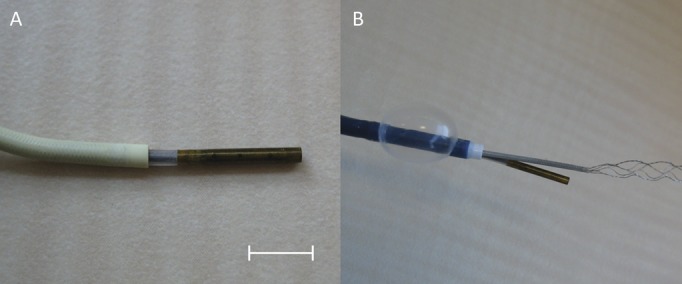
Background: Endoluminal optical imaging, or angioscopy, has not seen widespread application during neurointerventional procedures, largely as a result of the poor imaging resolution of existing angioscopes. Scanning fiber endoscopes (SFEs) are a novel endoscopic platform that allows high-resolution video imaging in an ultraminiature form factor that is compatible with currently used distal access endoluminal catheters.

Objective: To test the feasibility and potential utility of high-resolution angioscopy with an SFE during common endovascular neurosurgical procedures.

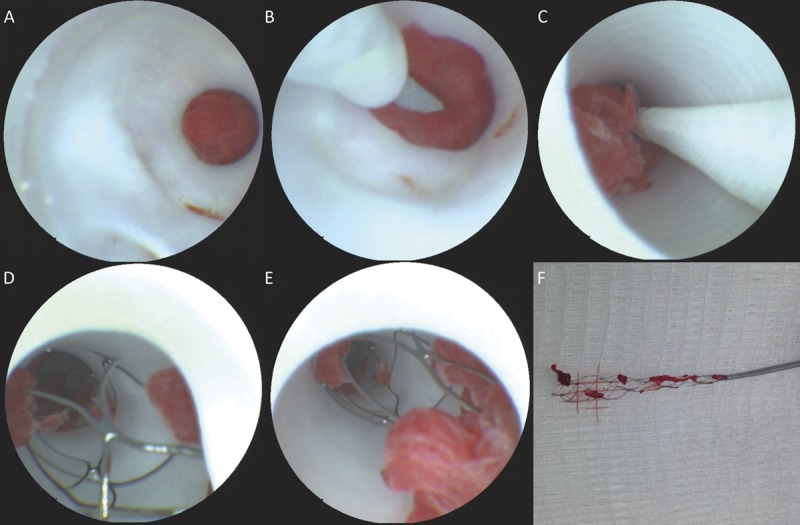
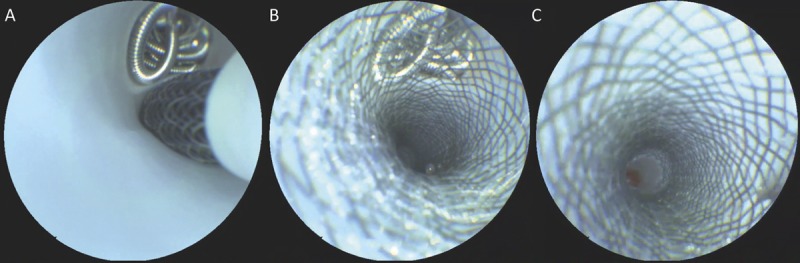
Methods: A 3.7-French SFE was used in a porcine model system to image endothelial disruption, ischemic stroke and mechanical thrombectomy, aneurysm coiling, and flow-diverting stent placement.

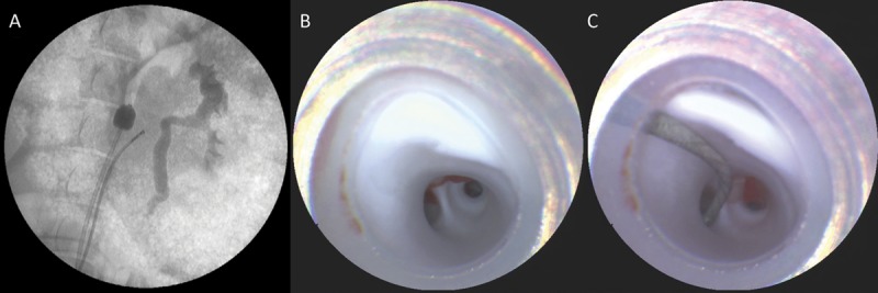
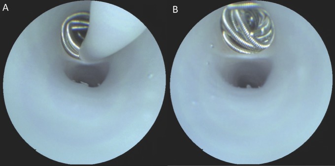
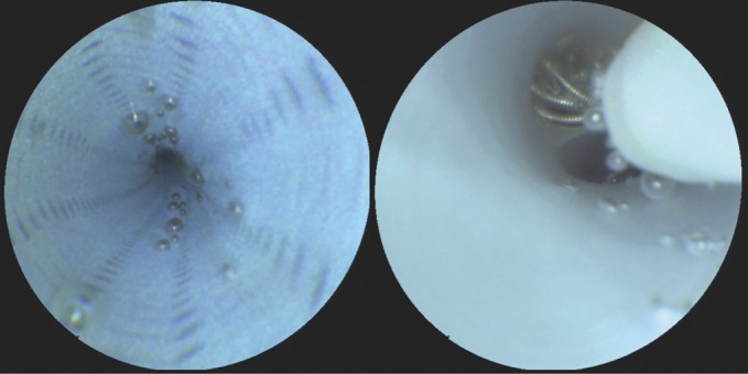
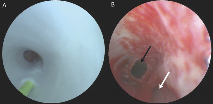
Results: High-resolution, video-rate imaging was shown to be possible during all of the common procedures tested and provided information that was complementary to standard fluoroscopic imaging. SFE angioscopy was able to assess novel factors such as aneurysm base coverage fraction and side branch patency, which have previously not been possible to determine with conventional angiography.

Conclusion: Endovascular imaging with an SFE provides important information on factors that cannot be assessed fluoroscopically and is a novel platform on which future neurointerventional techniques may be based because it allows for periprocedural inspection of the integrity of the vascular system and the deployed devices. In addition, it may be of diagnostic use for inspecting the vascular wall and postprocedure device evaluation.


Figures

FIGURE 1
FIGURE 1
A, a 3.7-French (F) scanning fiber endoscope (SFE) used in this study exiting from a 5-F Envoy guide catheter, demonstrating the 8-mm radiopaque tip. Scale bar = 5 mm. B, the SFE deployed in combination with a 9-F coaxial balloon guide catheter and 2.5-F microcatheter for imaging of mechanical thrombectomy with a stent retriever.
FIGURE 2
FIGURE 2
A, fluoroscopic image of the scanning fiber endoscope (SFE) in place in the renal artery with a balloon in the descending aorta for proximal flow control. B, view from the SFE at the end of the guide catheter, showing a smooth white endothelium and the first bifurcation of the renal artery. C, SFE image of renal branch selection using a 0.014-inch microwire.
FIGURE 3
FIGURE 3
Real-time scanning fiber endoscope images of aneurysm coil delivery (A) into a side branch from a microcatheter having a distal diameter of 2.8-French. B, post-filling inspection of the coil mass demonstrating a hanging detachment zone end and dense packing of coils at the level of the orificium.
FIGURE 4
FIGURE 4
Left, scanning fiber endoscope image demonstrating persistent microbubbles within the lumen of a fastidiously prepared guide catheter that were observed to dislodge randomly with increased saline solution flush rates. Right, image showing the displacement of microbubbles trapped within a flow-diverting stent during device deployment.
FIGURE 5
FIGURE 5
Scanning fiber endoscope images of ischemic stroke thrombectomy from initial lesion inspection (A), microwire crossing (B), initial withdrawal (C), inspection of stent retriever contents in situ (D, E), and ex vivo correlative image of retrieved material (F).
FIGURE 6
FIGURE 6
Scanning fiber endoscope imaging of flow-diverter placement over a previously coiled side branch showing placement of the stent during exit from the 2.8-French delivery microcatheter (A), the ability to resolve the number of diverter tines overlying the base of the coiled branch and any coil mass movement (B), and inspection of side-wall apposition, wall kinking, and side-branch patency (C).
FIGURE 7
FIGURE 7
Scanning fiber endoscope images of the development of an endothelial dissection. A, 0.014-inch microwire being applied in reverse showing the proximal entry point of the wire into the subendothelial space as well as the body of the wire tunneling distally. B, fully developed dissection with a damaged endothelium demonstrating the ability to differentiate the true (black arrow) and false (white arrow) lumens using angioscopy.

References

    1. Stonebridge P, Murie J. Angioscopy—a new light on peripheral vascular-disease. Eur J Vasc Surg. 1992;6(4):346-353 - PubMed
    1. Tokuhiro K, Uchida Y, Kawamura K, et al. Evaluation of annuloaortic ectasia by angioscopy and IVUS “report of 2 cases”. Diagn Ther Endosc. 2000;7(1):35-45 - PMC - PubMed
    1. Tsagakis K, Kamler M, Benedik J, Jakob H. Angioscopy—a valuable tool in guiding hybrid stent grafting and decision making during type A aortic dissection surgery. Eur J Cardiothoracic Surg. 2010;38(4):507-509 - PubMed
    1. Uchida Y. Recent advances in coronary angioscopy. J Cardiol. 2011;57(1):18-30 - PubMed
    1. Ishihara T, Iida O, Awata M, Nanto K, Nanto S, Uematsu M. Angioscopic assessment of early phase arterial repair after paclitaxel-coated nitinol drug-eluting stent implantation in the superficial femoral artery. Circ J. 2013;77(7):1838-1843 - PubMed

Publication types