. 2014 May 1;127(Pt 9):1857-67.
doi: 10.1242/jcs.113233. Epub 2014 Apr 24.

Nutrient-driven O-GlcNAc cycling - think globally but act locally

Affiliations

Nutrient-driven O-GlcNAc cycling - think globally but act locally

Katryn R Harwood et al. J Cell Sci. .

Abstract

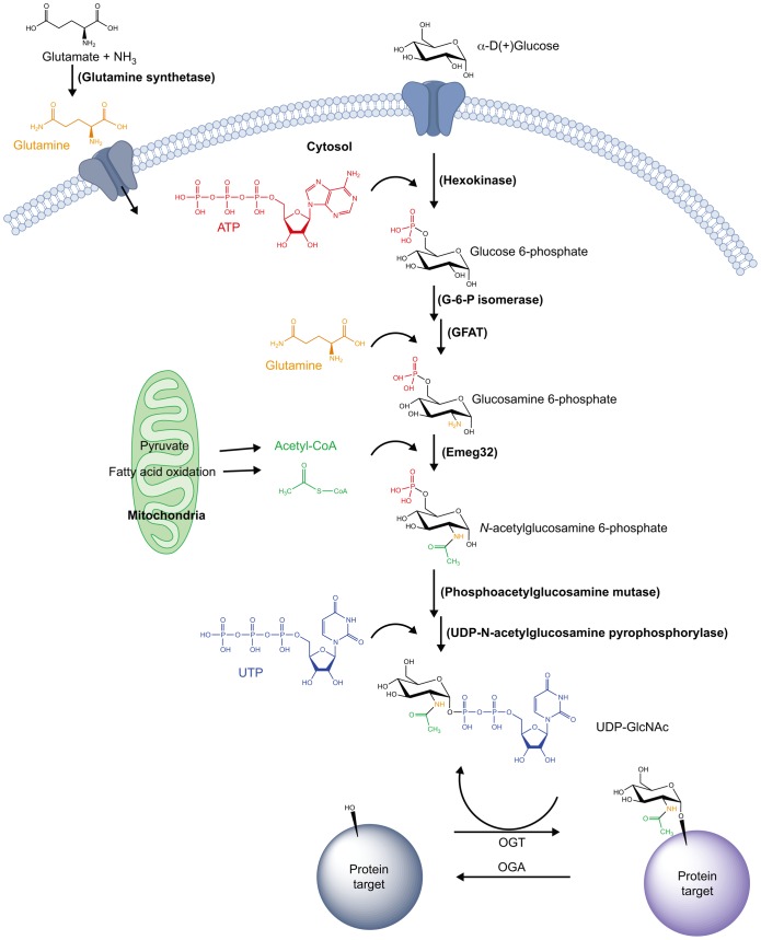
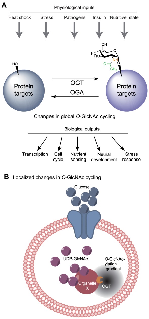
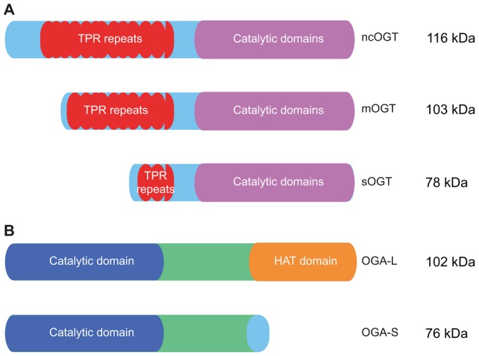
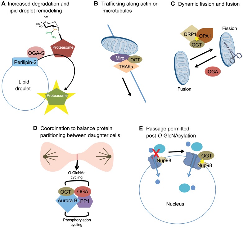
Proper cellular functioning requires that cellular machinery behave in a spatiotemporally regulated manner in response to global changes in nutrient availability. Mounting evidence suggests that one way this is achieved is through the establishment of physically defined gradients of O-GlcNAcylation (O-linked addition of N-acetylglucosamine to serine and threonine residues) and O-GlcNAc turnover. Because O-GlcNAcylation levels are dependent on the nutrient-responsive hexosamine signaling pathway, this modification is uniquely poised to inform upon the nutritive state of an organism. The enzymes responsible for O-GlcNAc addition and removal are encoded by a single pair of genes: both the O-GlcNAc transferase (OGT) and the O-GlcNAcase (OGA, also known as MGEA5) genes are alternatively spliced, producing protein variants that are targeted to discrete cellular locations where they must selectively recognize hundreds of protein substrates. Recent reports suggest that in addition to their catalytic functions, OGT and OGA use their multifunctional domains to anchor O-GlcNAc cycling to discrete intracellular sites, thus allowing them to establish gradients of deacetylase, kinase and phosphatase signaling activities. The localized signaling gradients established by targeted O-GlcNAc cycling influence many important cellular processes, including lipid droplet remodeling, mitochondrial functioning, epigenetic control of gene expression and proteostasis. As such, the tethering of the enzymes of O-GlcNAc cycling appears to play a role in ensuring proper spatiotemporal responses to global alterations in nutrient supply.

Keywords: Epigenetics; Lipid droplets; Metabolism; Mitochondria; Nuclear pores; O-GlcNAc; O-GlcNAcylation; Post-translational modification; Signaling.


Figures

Fig. 1.
Fig. 1.
Generation of the OGT substrate, UDP-GlcNAc – an overview of the hexosamine biosynthetic pathway. The hexosamine biosynthetic pathway (HBP) integrates many extracellular physiological inputs, including nutritional intake, with the metabolism of key carbohydrate, amino acid, nucleotide and fatty acid components, to produce the OGT substrate UDP-GlcNAc. O-GlcNAc cycling, the dynamic addition to and removal of O-GlcNAc on serine or threonine residues of a plethora of OGT and OGA targets, allows for the regulation of important downstream cellular processes in a nutrient-dependent manner. The enzymes involved are given in brackets.
Fig. 2.
Fig. 2.
Global and local responses to changes in UDP-GlcNAc levels. (A) The cell can respond to certain extracellular physiological inputs with global changes in intracellular UDP-GlcNAc levels and subsequent changes in total protein O-GlcNAcylation levels to modulate proper downstream cellular responses. (B) The tethering of OGA or OGT at specific cellular locations could establish local gradients of protein O-GlcNAcylation, and thus potentially introduce gradients of protein activity (illustrated by the gray ‘cloud’) that allow for highly specific spatiotemporal responses to the overall UDP-GlcNAc levels, with consequent cellular responses.
Fig. 3.
Fig. 3.
OGT and OGA isoforms and domain structure. (A) The three splice isoforms of OGT, ncOGT, mOGT and sOGT, possess variable N-termini but identical catalytic domains. (B) The two splice isoforms of OGA, OGA-L and OGA-S, differ at their C-termini. OGA-L possesses a putative HAT domain, whereas OGA-S lacks this domain and instead contains a unique 15-amino-acid long C-terminal extension.
Fig. 4.
Fig. 4.
O-GlcNAc cycling and localized cellular remodeling. (A) Lipid droplet remodeling. The targeted localization of OGA-S by perilipin-2 at the surface of lipid droplets results in the removal of O-GlcNAc from the proteasome and its subsequent activation. Proteasomal activation at the surface of lipid droplets increases the degradation of surface localized proteins, resulting in lipid droplet remodeling. (B) Mitochondrial trafficking. OGT binds to TRAKs and has a role in the trafficking of mitochondria along actin or microtubule networks, potentially either directly through TRAK binding, via TRAK O-GlcNAcylation, or both. (C) Mitochondrial fission and fusion. OGT O-GlcNAcylates DRP1 and OPA1 (indicated by the yellow star), which results in increased mitochondrial fission. Overexpression of OGA reverses this effect and promotes mitochondrial fusion. (D) Cytokinesis. During cytokinesis, both OGT and OGA are found in a transient complex with both Aurora B and the phosphatase PP1. It has been shown that the localization of OGT in this complex is dependent on Aurora B. These pairs of enzymes with opposing activities somehow coordinate their activities in a presumably complex manner to ensure proper protein partitioning into daughter cells and completion of cytokinesis. (E) Nuclear pore remodeling. O-GlcNAcylation of Nup98 by OGT (yellow star) can relax the ‘sieve’-like structure of the nuclear pore, allowing the passage of larger molecules not permitted prior to Nup98 O-GlcNAcylation.
Fig. 5.
Fig. 5.
O-GlcNAc cycling and transcriptional regulation. The proteins Sin3 (shown on the left) and TETs (shown on the right) might interact with OGT at targeted locations to repress or activate, respectively, the transcription of a specific subset of genes in response to nutritional cues. Simultaneously, the putative histone acetyl transferase (HAT) domain of OGA might regulate its binding to specific histone tail marks, allowing OGA, in conjunction with histone acetylation, to activate gene transcription.

Similar articles

Cited by

References

    1. Adams R. R., Maiato H., Earnshaw W. C., Carmena M. (2001). Essential roles of Drosophila inner centromere protein (INCENP) and aurora B in histone H3 phosphorylation, metaphase chromosome alignment, kinetochore disjunction, and chromosome segregation. J. Cell Biol. 153, 865–880 10.1083/jcb.153.4.865 - DOI - PMC - PubMed
    1. Bickel P. E., Tansey J. T., Welte M. A. (2009). PAT proteins, an ancient family of lipid droplet proteins that regulate cellular lipid stores. Biochim. Biophys. Acta 1791, 419–440 10.1016/j.bbalip.2009.04.002 - DOI - PMC - PubMed
    1. Blatch G. L., Lässle M. (1999). The tetratricopeptide repeat: a structural motif mediating protein-protein interactions. Bioessays 21, 932–939 10.1002/(SICI)1521--1878(199911)21:11<932::AID--BIES5>3.0.CO;2--N - DOI - PubMed
    1. Bond M. R., Hanover J. A. (2013). O-GlcNAc cycling: a link between metabolism and chronic disease. Annu. Rev. Nutr. 33, 205–229 10.1146/annurev--nutr--071812--161240 - DOI - PMC - PubMed
    1. Brasaemle D. L. (2007). Thematic review series: adipocyte biology. The perilipin family of structural lipid droplet proteins: stabilization of lipid droplets and control of lipolysis. J. Lipid Res. 48, 2547–2559 10.1194/jlr.R700014--JLR200 - DOI - PubMed

Publication types

MeSH terms

Substances