Aldehyde dehydrogenase 3A1 associates with prostate tumorigenesis
- PMID: 24762960
- PMCID: PMC4021532
- DOI: 10.1038/bjc.2014.201
Aldehyde dehydrogenase 3A1 associates with prostate tumorigenesis
Abstract
Background: Accumulating evidence demonstrates high levels of aldehyde dehydrogense (ALDH) activity in human cancer types, in part, because of its association with cancer stem cells. Whereas ALDH1A1 and ALDH7A1 isoforms were reported to associate with prostate tumorigenesis, whether other ALDH isoforms are associated with prostate cancer (PC) remains unclear.
Methods: ALDH3A1 expression was analysed in various PC cell lines. Xenograft tumours and 54 primary and metastatic PC tumours were stained using immunohistochemistry for ALDH3A1 expression.
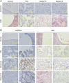
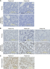
Results: In comparison with the non-stem counterparts, a robust upregulation of ALDH3A1 was observed in DU145-derived PC stem cells (PCSCs). As DU145 PCSCs produced xenograft tumours with more advanced features compared with those derived from DU145 cells, higher levels of ALDH3A1 were detected in the former; a dramatic elevation of ALDH3A1 occurred in DU145 cell-derived lung metastasis compared with local xenograft tumours. Furthermore, while ALDH3A1 was not observed in prostate glands, ALDH3A1 was clearly present in PIN, and further increased in carcinomas. In comparison with the paired local carcinomas, ALDH3A1 was upregulated in lymph node metastatic tumours; the presence of ALDH3A1 in bone metastatic PC was also demonstrated.
Conclusions: We report here the association of ALDH3A1 with PC progression.
Figures


Similar articles
-
IQGAP2, A candidate tumour suppressor of prostate tumorigenesis.Biochim Biophys Acta. 2012 Jun;1822(6):875-84. doi: 10.1016/j.bbadis.2012.02.019. Epub 2012 Mar 2. Biochim Biophys Acta. 2012. PMID: 22406297
-
Aldehyde dehydrogenase 3A1 is robustly upregulated in gastric cancer stem-like cells and associated with tumorigenesis.Int J Oncol. 2016 Aug;49(2):611-22. doi: 10.3892/ijo.2016.3551. Epub 2016 Jun 1. Int J Oncol. 2016. PMID: 27279633
-
Upregulation of FAM84B during prostate cancer progression.Oncotarget. 2017 Mar 21;8(12):19218-19235. doi: 10.18632/oncotarget.15168. Oncotarget. 2017. PMID: 28186973 Free PMC article.
-
Androgen receptor and prostate cancer stem cells: biological mechanisms and clinical implications.Endocr Relat Cancer. 2015 Dec;22(6):T209-20. doi: 10.1530/ERC-15-0217. Epub 2015 Aug 18. Endocr Relat Cancer. 2015. PMID: 26285606 Free PMC article. Review.
-
Stem cells in genetically-engineered mouse models of prostate cancer.Endocr Relat Cancer. 2015 Dec;22(6):T199-208. doi: 10.1530/ERC-15-0367. Epub 2015 Sep 4. Endocr Relat Cancer. 2015. PMID: 26341780 Free PMC article. Review.
Cited by
-
Targeting aldehyde dehydrogenase for prostate cancer therapies.Front Oncol. 2022 Oct 10;12:1006340. doi: 10.3389/fonc.2022.1006340. eCollection 2022. Front Oncol. 2022. PMID: 36300093 Free PMC article. Review.
-
Aldehyde dehydrogenase in solid tumors and other diseases: Potential biomarkers and therapeutic targets.MedComm (2020). 2023 Jan 16;4(1):e195. doi: 10.1002/mco2.195. eCollection 2023 Feb. MedComm (2020). 2023. PMID: 36694633 Free PMC article. Review.
-
Silencing of NRF2 Reduces the Expression of ALDH1A1 and ALDH3A1 and Sensitizes to 5-FU in Pancreatic Cancer Cells.Antioxidants (Basel). 2017 Jul 1;6(3):52. doi: 10.3390/antiox6030052. Antioxidants (Basel). 2017. PMID: 28671577 Free PMC article.
-
Identification of a biomarker panel for improvement of prostate cancer diagnosis by volatile metabolic profiling of urine.Br J Cancer. 2019 Nov;121(10):857-868. doi: 10.1038/s41416-019-0585-4. Epub 2019 Oct 7. Br J Cancer. 2019. PMID: 31588123 Free PMC article.
-
Expansion of mouse castration-resistant intermediate prostate stem cells in vitro.Stem Cell Res Ther. 2022 Jul 15;13(1):299. doi: 10.1186/s13287-022-02978-x. Stem Cell Res Ther. 2022. PMID: 35841025 Free PMC article.
References
-
- Bonnet D, Dick J. Human acute myeloid leukemia is organized as a hierarchy that originates from a primitive hematopoietic cell. Nat Med. 1997;7:730–737. - PubMed
-
- Brabletz T, Jung A, Spaderna S, Hlubek F, Kirchner T. Opinion: migrating cancer stem cells - an integrated concept of malignant tumour progression. Nat Rev Cancer. 2005;5 (9:744–749. - PubMed
-
- Canuto R, Muzio G, Ferro M, Maggiora M, Federa R, Bass A, Lindahl R, Dianzani M. Inhibition of class 3 aldehyde dehydrogenase and cell growth by restored lipid peroxidation in heptoma cell lines. Free Radic Biol Med. 1999;26:333–340. - PubMed
-
- Charafe-Jauffret E, Ginestier C, Iovino F, Tarpin C, Diebel M, Esterni B, Houvenaeghel G, Extra J-M, Bertucci F, Jacquemier J, Xerri L, Dontu G, Stassi G, Xiao Y, Barsky SH, Birnbaum D, Viens P, Wicha MS. Aldehyde dehydrogenase 1-positive cancer stem cells mediate metastasis and poor clinical outcome in inflammatory breast cancer. Clin Cancer Res. 2010;16:45–55. - PMC - PubMed
Publication types
MeSH terms
Substances
Grants and funding
LinkOut - more resources
Full Text Sources
Other Literature Sources
Medical
Research Materials
Miscellaneous

