. 2014;13(12):1970-9.
doi: 10.4161/cc.28922. Epub 2014 Apr 24.

Mammalian cells contain two functionally distinct PBAF complexes incorporating different isoforms of PHF10 signature subunit

Affiliations

Mammalian cells contain two functionally distinct PBAF complexes incorporating different isoforms of PHF10 signature subunit

Alexander V Brechalov et al. Cell Cycle. 2014.

Abstract

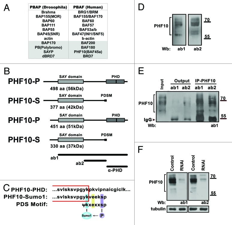
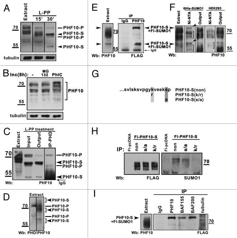
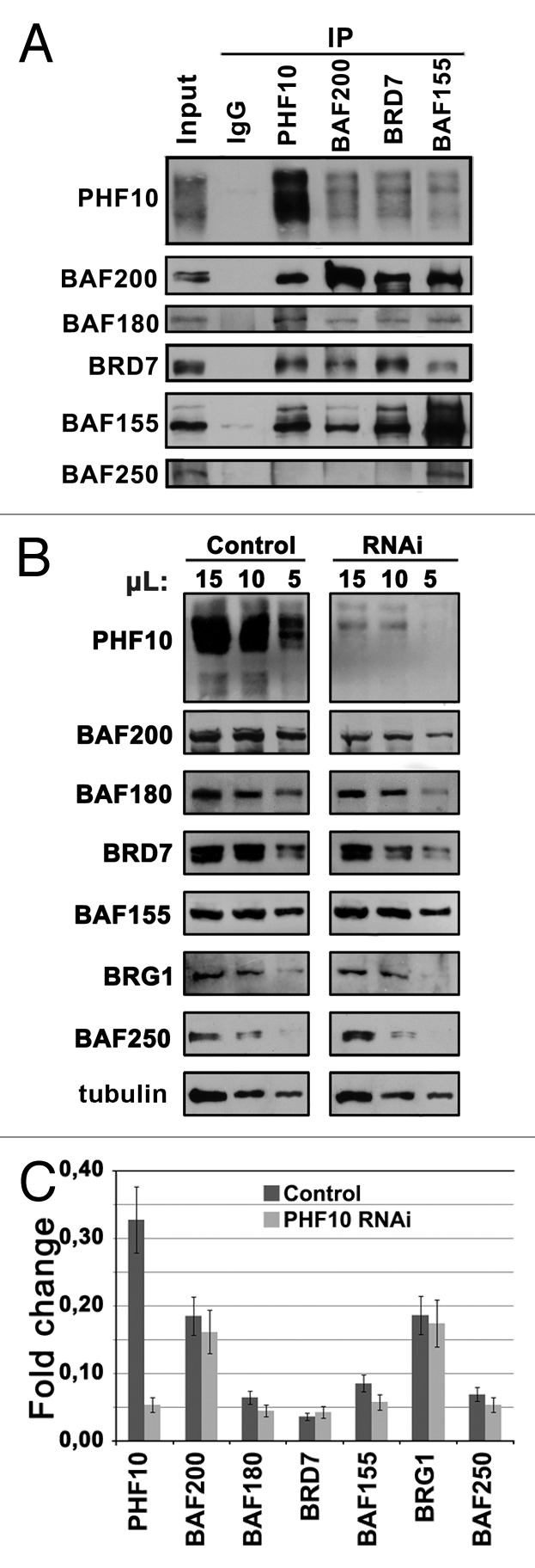
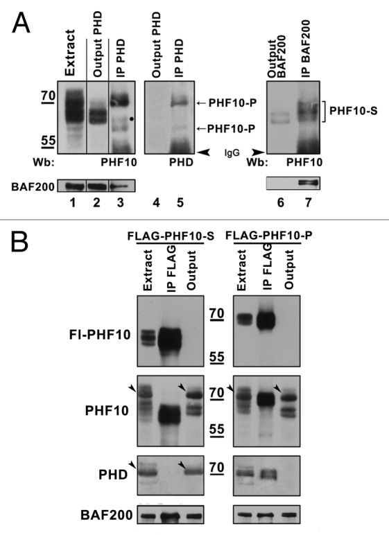
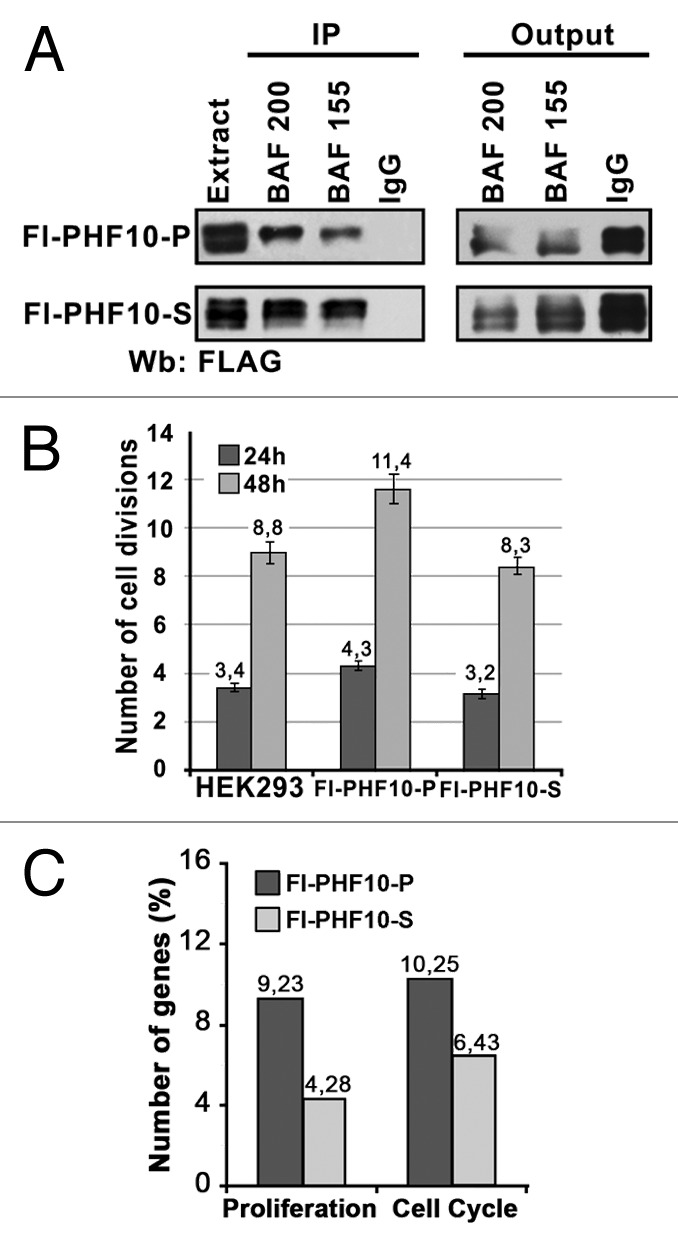
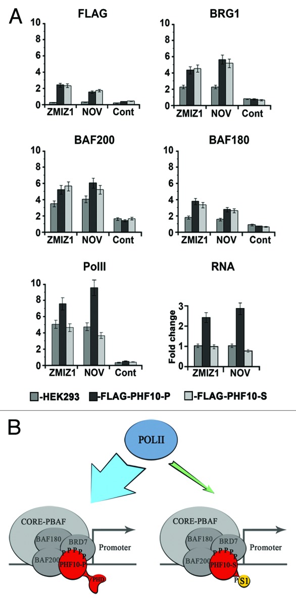
The PBAF subtype of the mammalian chromatin remodeling SWI/SNF complex has wide and diverse functions in transcription regulation and development, being both transcription activator and repressor. However, a mechanism accounting for such functional diversity remains unclear. Human PHF10/BAF45a subunit of the PBAF complex plays an important role in brain development but has not been studied sufficiently. We have shown that the PHF10 gene encodes 2 types of evolutionarily conserved, ubiquitously expressed isoforms that are incorporated into the PBAF complex in a mutually exclusive manner. One isoform contains C-terminal tandem PHD fingers, which in the other isoform are replaced by the consensus sequence for phosphorylation-dependent SUMO 1 conjugation (PDSM). PBAF complexes containing different PHF10 isoforms can bind to the promoters of the same genes but produce different effects on the recruitment of Pol II to the promoter and on the level of gene transcription. In addition, it is only the PBAF with PHD-containing isoform that activates proliferation. Our study demonstrates the existence of functionally different PBAF complexes in mammalian cell. It also provides an insight into the molecular structure and role of human PHF10/BAF45a and characterizes it as an essential PBAF subunit.

Keywords: PBAF signature subunits; PDSM motif; PHD domains; PHF10/BAF45; SWI/SNF chromatin remodeling complex; functionally distinct isoforms; sumoylation.


Figures

None
Figure 1. The PHF10 gene encodes isoforms containing either PHD fingers or PDSM motif as the C-terminal domain. (A) The subunit composition of Drosophila and mammalian PBAF complexes. The homologous subunits are grouped horizontally. The PBAF-specific subunits are shown on a gray background. (B) The predicted structure of PHF10 isoforms. The PHF10 mRNA encodes isoforms that have an evolutionary conserved SAY domain and tandem PHD domains (PHF10-P) or PDSM motif (PHF10-S) at the C terminus. The short PHF10-P and PHF10-S lack 47 N-terminal amino acids. The full-length PHF10-P isoform corresponds to the PHF10 protein annotated earlier (NP_060758.2). The regions used to raise antibodies are shown at the bottom. (C) The consensus sequence for phosphorylation-dependent SUMO modification (PDSM) at the C terminus of PHF10-S isoforms. Sequences common to PHF10-P and PHF10-S are boxed. (D) Detection of PHF10 in HEK293 cell extract by western blotting with 2 different affinity purified anti-PHF10 antibodies (ab1 and ab2). (E) Each of anti-PHF10 antibodies precipitates PHF10 bands from the HEK293 extract. (F) The shRNAi knockdown of PHF10 leads to depletion of PHF10 isoforms from the HEK293 extract.
None
Figure 2. Post-translational modifications of PHF10 isoforms. (A) Treatment of HEK293 cell extract with lambda protein phosphatase (L-PP) for 15 or 30 min leads to reduction of numerous PHF10 isoforms to 4 stable protein bands (arrowheads). (B) Inhibition of phosphatases stabilizes high-molecular-weight PHF10 bands. Western blotting of PHF10 in protein extracts from wild-type HEK293 cells and from HEK293 cells grown for 8 h in the presence of MG132 proteasome inhibitor or phosphatase inhibitor cocktail (PhIC). Accumulation of high-molecular-weight PHF10 bands (arrowheads) can be seen in cells grown in the presence of PhIC. (C) Immunoprecipitation of PHD-containing PHF10 forms (PHF10-P) performed with anti-PHD antibodies from the HEK293 extract treated with L-PP. The PHF10-S isoforms remain in the output material. (D) Western blotting of HEK293 extract with anti-PHD or anti-PHF10 (ab1) antibodies. The PHF10-P and PHF10-S isoforms are indicated. (E) Endogenous PHF10-S isoforms modified by SUMO 1 conjugation. FLAG-tagged SUMO 1 was transiently expressed in HEK293 cells, and the cell extract was immunoprecipitated by anti-PHF10 antibodies. Western blotting of the precipitate with anti-FLAG antibodies revealed 2 sumoylated PHF10 forms. Their electrophoretic mobility coincided with that of modified PHF-S isoforms in the HEK293 extract (left panel, indicated by arrowheads). (F) Nuclear extract from HEK293 cells transiently transfected with 10 His- SUMO 1 or from control (nontransfected) HEK293 cells was incubated with Ni-NTA, and precipitated proteins were resolved by western blotting with antibodies against PHF10. Sumoylated PHF10 bound by Ni-NTA resin (arrows) was depleted from the pool of PHF10 isoforms (see “Output”). The PHF10 forms indicated with brackets are those nonspecifically bound by Ni-NTA. (G) The amino acid sequence of wild-type and mutated PDSM. In mutants, either serine was replaced by alanine or lysine was replaced by arginine to prevent phosphorylation or sumoylation, respectively. (H) The PDSM motif of PHF10-S conjugates SUMO 1 in a phosphorylation-dependent manner. FLAG-tagged wild-type and mutated forms of PHF10-S were transiently expressed HEK293 cells and purified on anti-FLAG agarose. The recombinant PHF10 was precipitated with anti-FLAG antibodies from the cell extract, and equal amounts of precipitate were loaded onto a gel (FLAG). Staining with anti-SUMO 1 antibody revealed sumoylated recombinant PHF10 (SUMO 1). (I) The sumoylated PHF10-S is associated with PBAF. Immunoprecipitation with antibodies against BAF200 or BAF155 was performed from the extract of HEK293 cells stably transfected with FLAG-tagged SUMO 1. The western blot was developed with antibodies against FLAG to detect sumoylated PHF10 (arrowhead). Immunoprecipitation with antibodies against tubulin was used as a negative control.
None
Figure 3. PHF10 isoforms are associated with PBAF signature subunits. (A) Immunoprecipitation (IP) of PBAF from HEK293 extract with antibodies against PHF10 (ab1), BAF200, and BRD7 specific PBAF subunits and a BAF155 core SWI/SNF subunit. (B) The RNAi knockdown of PHF10 leads to a decrease in the level of specific PBAF subunits in the cells. HEK293 cells were stably transfected with the construct expressing shRNA for PHF10 RNAi or with an empty vector (control). The equal amounts of protein extracts from PHF10 RNAi or control cells were loaded onto the gel, and the western blot was developed with antibodies against PBAF subunits. Tubulin was used as a loading control. (C) Transcription levels of different PBAF subunits against the background of PHF10 RNAi and in control cells as measured by qPCR.
None
Figure 4. PHF10 isoforms containing PHD fingers or PDSM motif are associated with distinct PBAF complexes. (A) PBAF complexes containing distinct PHF10 isoforms are present in the HEK293 cell extract. The PHF10-P associated PBAF complexes were depleted from the extract with anti-PHD antibodies (lanes 1–5), while the complexes that contained PHF10-S remained in the output material and were further precipitated with anti-BAF200 antibodies (lanes 6–7). The western blot was developed with antibodies against PHF10 (lanes 1–3, 6, 7) or anti-PHD (lanes 4, 5). A significant amount of BAF200 co-precipitated with each of the PHF10 isoforms (lower panel), suggesting that PBAF complexes with PHF10-P or PHF10-S were both well represented in the cells. (B) Recombinant FLAG-tagged PHF10-S (left panel) or PHF10-P (right panel) in stably transfected HEK293 cells do not co-precipitate with endogenous PHF10-P and PHF10-Sl isoforms. The western blot was developed with anti-FLAG, anti-PHF10, and anti-PHD antibodies. Each recombinant isoform (panel FLAG-PHF10) was completely depleted from the extract, while the endogenous isoform with a different C-terminal domain remained in the output material (panels PHF10, PHD). Endogenous isoforms PHF10-P (left panel) and PHF10-Sl (right panel) are indicated by arrowheads.
None
Figure 5. PHF10 isoforms containing PHD fingers or PDSM motif have different effects on cell proliferation. (A) Recombinant PHF10-P and PHF10-S isoforms are associated with PBAF. Immunoprecipitation with antibodies against BAF200 or BAF155 was performed from the extract of HEK293 cells stably transfected with FLAG-tagged PHF10-P or PHF10-S. The western blot was developed with antibodies against FLAG to detect recombinant PHF10. (B) Numbers of cell divisions recorded 24 and 48 h after plating in a wild-type HEK293 culture and in cells stably transfected with FLAG-PHF10-P or FLAG- PHF10-S. Cells were stained with CellTrace Violet Kit and counted in a flow cytometer. The proportion of necrotic cells (propidium iodide staining) in any sample did not exceed 5%. (C) The results of gene expression analysis with Illumuna HumanHT-12 microarray. The percentages of genes involved in proliferation and cell cycle (DAVID Functional Annotation Tool) relative to the total number of differentially expressed genes are shown for FLAG-PHF10-P (black bars) or FLAG- PHF10-S (gray bars) transfected cells.
None
Figure 6. PHF10-P and PHF10-S have different effects on Pol II recruitment to promoter and on transcription level. (A) The presence of FLAG-PHF10-P, FLAG-PHF10-S, PBAF subunits, and Pol II on the promoters of ZMIZ1 and NOV genes and their transcription level (RNA) in HEK293 and stably transfected cells as determined by ChIP. The intergenic region was used as a control. (C) Two types of PBAF complexes that contain PHF10 with either PHD fingers or a PDSM motif at the C terminus are present in human cells. PHF10 is an intrinsic subunit of the PBAF signature module and is stably associated with BAF200 and BAF155. Both types of PHF10 isoforms are phosphorylated, and the PDSM isoform is also sumoylated. The isoform containing the PHD fingers, but not that with the PDSM motif, facilitates the recruitment of Pol II to the promoter and enhances the transcription level of the gene.

Similar articles

Cited by

References

    1. Wu JI. Diverse functions of ATP-dependent chromatin remodeling complexes in development and cancer. Acta Biochim Biophys Sin (Shanghai) 2012;44:54–69. doi: 10.1093/abbs/gmr099. - DOI - PubMed
    1. Mohrmann L, Verrijzer CP. Composition and functional specificity of SWI2/SNF2 class chromatin remodeling complexes. Biochim Biophys Acta. 2005;1681:59–73. doi: 10.1016/j.bbaexp.2004.10.005. - DOI - PubMed
    1. Kaeser MD, Aslanian A, Dong MQ, Yates JR, 3rd, Emerson BM. BRD7, a novel PBAF-specific SWI/SNF subunit, is required for target gene activation and repression in embryonic stem cells. J Biol Chem. 2008;283:32254–63. doi: 10.1074/jbc.M806061200. - DOI - PMC - PubMed
    1. Moshkin YM, Chalkley GE, Kan TW, Reddy BA, Ozgur Z, van Ijcken WF, Dekkers DH, Demmers JA, Travers AA, Verrijzer CP. Remodelers organize cellular chromatin by counteracting intrinsic histone-DNA sequence preferences in a class-specific manner. Mol Cell Biol. 2012;32:675–88. doi: 10.1128/MCB.06365-11. - DOI - PMC - PubMed
    1. Bultman S, Gebuhr T, Yee D, La Mantia C, Nicholson J, Gilliam A, Randazzo F, Metzger D, Chambon P, Crabtree G, et al. A Brg1 null mutation in the mouse reveals functional differences among mammalian SWI/SNF complexes. Mol Cell. 2000;6:1287–95. doi: 10.1016/S1097-2765(00)00127-1. - DOI - PubMed

Publication types

LinkOut - more resources