Review
. 2014 Apr 25;114(9):1500-15.
doi: 10.1161/CIRCRESAHA.114.303772.

Role of the autonomic nervous system in atrial fibrillation: pathophysiology and therapy

Affiliations
Review

Role of the autonomic nervous system in atrial fibrillation: pathophysiology and therapy

Peng-Sheng Chen et al. Circ Res. .

Abstract

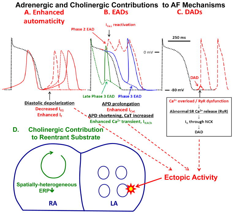
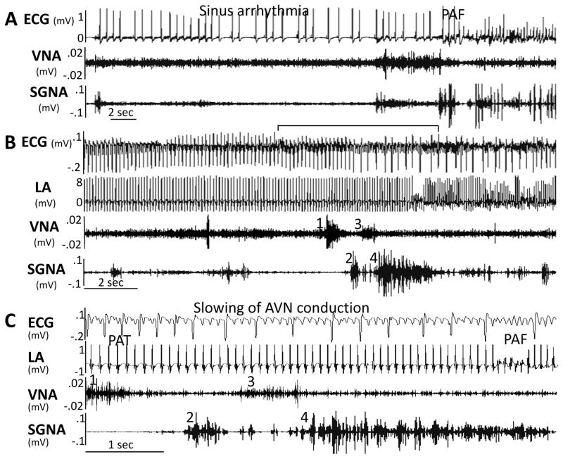
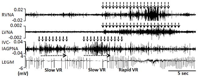
Autonomic nervous system activation can induce significant and heterogeneous changes of atrial electrophysiology and induce atrial tachyarrhythmias, including atrial tachycardia and atrial fibrillation (AF). The importance of the autonomic nervous system in atrial arrhythmogenesis is also supported by circadian variation in the incidence of symptomatic AF in humans. Methods that reduce autonomic innervation or outflow have been shown to reduce the incidence of spontaneous or induced atrial arrhythmias, suggesting that neuromodulation may be helpful in controlling AF. In this review, we focus on the relationship between the autonomic nervous system and the pathophysiology of AF and the potential benefit and limitations of neuromodulation in the management of this arrhythmia. We conclude that autonomic nerve activity plays an important role in the initiation and maintenance of AF, and modulating autonomic nerve function may contribute to AF control. Potential therapeutic applications include ganglionated plexus ablation, renal sympathetic denervation, cervical vagal nerve stimulation, baroreflex stimulation, cutaneous stimulation, novel drug approaches, and biological therapies. Although the role of the autonomic nervous system has long been recognized, new science and new technologies promise exciting prospects for the future.

Keywords: heart failure; myocardial infarction.


Figures

Figure 1
Figure 1
Autonomic innervation and neuromodulation. VLCCN, ventral lateral cervical cardiac nerve; VMCCN, ventromedial cervical cardiac nerve. Neiguan P6 is an acupoint used in a clinical trial of AF. The black dots indicate sites used by various investigators for neuromodulation to control AF. See Neuromodulation section for details. Illustration Credit: Ben Smith.
Figure 2
Figure 2
Presence of both adrenergic and cholinergic nerves structures in the extrinsic cardiac nervous system. Panel A is a low power view of the left stellate ganglion, showing numerous ganglion cells and nerve fibers stained positively for tyrosine hydroxylase. While most of the ganglion cells are tyrosine hydroxylase (TH) positive, some ganglion cells were negative (Panel B). Panels C shows tyrosine hydroxylase staining of a different stellate ganglion, showing tyrosine hydroxylase-negative cells (arrows). These same cells stained positive for cholineacetyltransferase (ChAT, arrows in Panel D). Some cells stain positive for both markers. These figures came from Shen et al, with permission. Panels E and F show tyrosine hydroxylase and cholineacetylesterase stains, respectively, of the canine left cervical vagal nerve. Arrows point to cells that stained positive for both markers. From Onkka et al, with permission.
Figure 3
Figure 3
Tyrosine hydroxylase (TH) and cholineacetyltransferase (ChAT) staining of the cervical vagal nerves. A: A low power view of the right cervical vagal nerve stained with tyrosine hydroxylase. There are 2 distinct nerve bundles in this nerve. B, D: The tyrosine hydroxylase stain of the smaller (B) and the larger (D) bundles in A. The brown color identifies the positively stained nerves. Note that tyrosine hydroxylase -positive nerves are located in the periphery of the nerve bundle. C, E: cholineacetyltransferase staining of the same structures as in panels B and D, respectively. Note that cholineacetyltransferase-positive components are widely distributed in the cervical vagal nerve. F: The tyrosine hydroxylase -positive nerve structure (red arrow) in the middle of the cervical vagal nerve. The objective lens used in panel A was 4X, with a calibration bar of 0.2 mm in length. The objective lens used in panels B–F was 20X, with a calibration bar of 0.2 mm in length. G shows the activation of vagal nerve alone is associated increased heart rate, a finding consistent with the activation of the sympathetic component of the vagal nerve. From Onkka et al, with permission.
Figure 4
Figure 4
Molecular basis for autonomic contributions to AF substrate. Beta-adrenergic receptor (β-AR) activation causes GTP-binding to the Gαs-subunite, allowing it to dissociate from Gβ and γ subunits and activate adenylate cyclase (AC), which converts ATP to cyclic-AMP (cAMP). cAMP activates protein-kinase A (PKA), which phosphorylates a range of Ca2+-handling proteins including the L-type Ca2+-channel (LTCC), ryanodine-receptor (RyR2) and phospholamban (PLB). PLB-phosphorylation causes it to dissociate from the sarcoplasmic-reticulum (SR) Ca2+-ATPase, SERCA2a, removing SERCA2a from PLB-inhibition and activating SR Ca2+-uptake. RyR2-phosphorylation increases RyR2 open probability, enhancing the systolic Ca2+-transient but also enhancing diastolic Ca2+-leak. Adrenergic stimulation also increases Ca2+ binding to calmodulin (CaM), activating Ca2+/CaM-dependent kinase type-II (CaMKII), which phosphorylates many of the same proteins as PKA. Ca2+/CaM also activates calcineurin (Cn), which dephosphorylates nuclear factor of activated T-cells (NFAT), allowing it to translocate to the nucleus and activate hypertrophic and profibrotic gene-programs. LTCC-phosphorylation increases ICaL and shifts its voltage-dependence to cause larger window-currents. Adrenergic stimulation also inhibits inward-rectifier K+-current (IK1) and enhances slow delayed-rectifier K+-current (IKs). Cholinergic activation of muscarinic type-2 (M2) acetylcholine-receptors (AChRs) causing GTP-binding to Gαi, releasing Gβγ and allowing it to activate the acetylcholine-dependent K+-current (IKACh).
Figure 5
Figure 5
Mechanisms by which autonomic tone can promote AF. Top: Action potential changes showing cellular mechanisms by which adrenergic activation can lead to focal ectopic firing. Black dotted tracings represent normal reference action potentials in each panel. A. Enhanced automaticity. B. Early afterdepolarizations (EADs). C. Delayed afterdepolarization (DADs). Contributions from adrenergic activation alone are shown by red tracings, while that from cholinergic activation (combined with adrenergic activation) by green tracings. Adrenergic stimulation in the setting of impaired repolarization reserve can cause phase-2 EADs (red dashed tracings in B). Most phase 3 EADs are also associated with prolonged APD (blue dashed tracings in B). Combined adrenergic/vagal discharge can produce late phase-3 EADs (green dashed tracings in B) due to a prolonged and enhanced Ca2+-transient that outlasts IKACh-induced accelerated repolarization. Bottom: Tissue-level arrhythmia mechanisms, with focal ectopic activity maintaining AF as a driver or acting on vulnerable reentrant substrates. Parasympathetic firing discharges acetylcholine, producing spatially-heterogeneous action-potential and refractory-period abbreviation that promotes the occurrence and maintenance of reentrant activity.
Figure 6
Figure 6
Two examples of paroxysmal atrial fibrillation (PAF). (A) Sinus rhythm to AF conversion. (B) Atrial tachycardia to AF conversion. (C) Magnified from the center of Panel B (line segment above ECG), showing that the elevated vagal nerve activity (VNA) accelerated atrial rate, leading to paroxysmal reduction of ventricular rate (prolonged RR interval) before conversion from paroxysmal atrial tachycardia (PAT) to paroxysmal atrial fibrillation (PAF). From Tan et al, with permission.
Figure 7
Figure 7
Local control of atrioventricular (AV) node conduction during persistent atrial fibrillation (AF). Slowing of ventricular rate (VR) was associated with inferior vena cava-inferior atrial ganglionated plexus nerve activity (IVC-IAGPNA) without either right vagal nerve activity (RVNA) or left vagal nerve activity (LVNA). Subsequent simultaneous activation of right vagal nerve activity and left vagal nerve activity resulted in a rapid ventricular rate. Because of the presence of abundant sympathetic nerves within the vagus, these observations suggest that sympathetic component within the vagal nerves have accelerated the ventricular rate. LEGM is the bipolar local electrogram showing ventricular activation. From Park et al, with permission.
Figure 8
Figure 8
Changes of type 2 small conductance calcium activated K (SK2) protein in the left stellate ganglion (LSG) with low level vagal nerve stimulation (VNS). A, Representative western blots show that the signal ratio of SK2 protein to GAPDH of vagal nerve stimulation dogs (Group 1) was significantly higher than that of control (Group 2). B, There is an upregulation of SK2 protein level in the LSG in Group 1 dogs after being normalized to GAPDH. C–D, Representative immunostaining of SK2 protein in the left stellate ganglion. The density of SK2-positive nerve structures (as pointed by a black arrowhead) is significantly higher in Group 1 dogs (Panel C), compared to Group 2 dogs (Panel D). E–F, Representative low-power view of immunostaining of SK2 protein in the LSG, that clearly demonstrates higher SK2 density in Group 1 dogs (Panel E), compared to Group 2 dogs (Panel F). G and H show immunofluorescence confocal microscope images of the LSG from Group 1 and Group 2 dogs, respectively. Blue colored dots show the nuclei stained with 4′,6-diamidino-2-phenylindole. Red color marks the SK2 protein. Note a significantly increased SK2 staining in the periphery of ganglion cells but decrease in the cytosol of Group 1 (G). In contrast, in Group 2 LSG, the SK2 staining was homogeneous (H). SK2, Small conductance calcium-activated potassium channels subtype 2; GAPDH, glyceraldehydes-3-phosphate-dehydrogenase; AU, arbitrary units. Calibration bar = 50 μm for C–F and 20 μM for G and H.

References

    1. Viskin S, Golovner M, Malov N, Fish R, Alroy I, Vila Y, Laniado S, Kaplinsky E, Roth A. Circadian variation of symptomatic paroxysmal atrial fibrillation. Data from almost 10 000 episodes. European Heart Journal. 1999;20:1429–1434. - PubMed
    1. Leiria TL, Glavinovic T, Armour JA, Cardinal R, de Lima GG, Kus T. Longterm effects of cardiac mediastinal nerve cryoablation on neural inducibility of atrial fibrillation in canines. Autonomic neuroscience: basic & clinical. 2011;161:68–74. - PubMed
    1. Richer LP, Vinet A, Kus T, Cardinal R, Ardell JL, Armour JA. Alpha-adrenoceptor blockade modifies neurally induced atrial arrhythmias. American journal of physiology. Regulatory, integrative and comparative physiology. 2008;295:R1175–1180. - PMC - PubMed
    1. Tan AY, Zhou S, Ogawa M, Song J, Chu M, Li H, Fishbein MC, Lin SF, Chen LS, Chen PS. Neural mechanisms of paroxysmal atrial fibrillation and paroxysmal atrial tachycardia in ambulatory canines. Circulation. 2008;118:916–925. - PMC - PubMed
    1. Shen MJ, Shinohara T, Park HW, Frick K, Ice DS, Choi EK, Han S, Maruyama M, Sharma R, Shen C, Fishbein MC, Chen LS, Lopshire JC, Zipes DP, Lin SF, Chen PS. Continuous low-level vagus nerve stimulation reduces stellate ganglion nerve activity and paroxysmal atrial tachyarrhythmias in ambulatory canines. Circulation. 2011;123:2204–2212. - PMC - PubMed

Publication types

Substances