Review
. 2014 Apr 25;114(9):1516-31.
doi: 10.1161/CIRCRESAHA.114.302240.

Mathematical approaches to understanding and imaging atrial fibrillation: significance for mechanisms and management

Affiliations
Review

Mathematical approaches to understanding and imaging atrial fibrillation: significance for mechanisms and management

Natalia A Trayanova. Circ Res. .

Abstract

Atrial fibrillation (AF) is the most common sustained arrhythmia in humans. The mechanisms that govern AF initiation and persistence are highly complex, of dynamic nature, and involve interactions across multiple temporal and spatial scales in the atria. This article aims to review the mathematical modeling and computer simulation approaches to understanding AF mechanisms and aiding in its management. Various atrial modeling approaches are presented, with descriptions of the methodological basis and advancements in both lower-dimensional and realistic geometry models. A review of the most significant mechanistic insights made by atrial simulations is provided. The article showcases the contributions that atrial modeling and simulation have made not only to our understanding of the pathophysiology of atrial arrhythmias, but also to the development of AF management approaches. A summary of the future developments envisioned for the field of atrial simulation and modeling is also presented. The review contends that computational models of the atria assembled with data from clinical imaging modalities that incorporate electrophysiological and structural remodeling could become a first line of screening for new AF therapies and approaches, new diagnostic developments, and new methods for arrhythmia prevention.

Keywords: arrhythmias, cardiac; atrial fibrillation; atrial remodeling; computer simulation.


Figures

Figure 1
Figure 1
A. Schematic of the atrial cell, including representations of ionic channels and intracellular ion concentrations used in the various human atrial cell models. B. Schematic of Ca handling with different compartments and currents in the various atrial cell models. Each of the models’ Ca handling is represented in one quarter of the schematic. White spaces in the central region denote the SR, with straight broken lines representing the division between SR uptake and release regions. Gray spaces represent the other compartments within the cell and outside of SR, with the “cleft” and “dyad” sub-spaces shown in white. Arrows indicate directions of Ca flow between the different compartments. The SR Ca pump is indicated by circle and arrow. The Koivumaki et al model also incorporates additional compartments representing peripheral and central SR regions and the current flow between these regions and the intracellular space. Additional detail regarding Ca handling in the models can be found in the original publications.,,,, D–G. Control and cAF APs of five atrial cell models paced at a frequency of 1Hz. Modified with permission from.
Figure 2
Figure 2
Geometrical models of the atria. A: Volume image of the sheep atria acquired by serial surface imaging (resolution 50 microns), with a representative slice. Subdivision of atria into different regions as represented by the different colors: RA—green, LA—blue, Bachman’s bundle (BB)—red, posterior left atrium (PLA)—yellow. Images reproduced with permission from,. B: A model of the fibrotic human atria generated from a patient LGE-MRI scan (top left) following segmentation (top right) into normal and fibrotic tissue (fibrotic lesions in red). With permission from.
Figure 3
Figure 3
Membrane potential distribution on the epicardial surface of the human atria. A: Spontaneous normal rhythm. B: Pacing-induced AF resulting from different electrophysiological properties of the crista terminalis. With permission from.
Figure 4
Figure 4
A: Continuous PV re-activation of the LA due to heterogeneous venous conduction and reentry following a single ectopic beat in the LA (with permission from). The sinus beat propagates heterogeneously along the vein (wrapped and unwrapped views, 342ms). Vein length is 1 cm, and circumference is 2cm, with 30% longitudinal and 65% transverse cellular disconnections. A single premature activation originating somewhere in LA invades PV following the sinus beat (342ms and 408ms), encountering block (408ms) and establishing PV reentry (492ms). This reentry continuously re-excites the LA, serving as “focal source” for LA activations (576ms; propagation entering LA at bottom of image). B: A snapshot of membrane voltage in the human atria at a single time point during AF (different views of the atria are shown), in which numbered arrows (1–4) indicate multiple reentrant wavelets. AF was induced by PV ectopic beats (with permission from). SVC and IVC–superior and inferior vena cava; BB--Bachman’s bundle; FW–free wall; TA—tricuspid annulus.
Figure 5
Figure 5
A: Simulated APs in control and chronic AF conditions (CAF1, CAF2). CAF1 is an AF cell model with Ito and ICaL reduced, without IK1 upregulation; CAF2 is the same model with IK1 increased. B: Electrical restitution plotted as APD–70 versus the diastolic interval (DI) in control and chronic AF cases. C and D: Spiral waves (phase movie snapshots) and tip meander in chronic AF conditions CAF1 and CAF2. Phase movies are shown at four distinct times. The figure demonstrates that IK1 stabilizes and accelerates reentry, as manifested by the reduced tip meander. Modified with permission from.
Figure 6
Figure 6
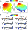
Modeling fibrosis as regions of collagen presence. Collagen is represented as an insulator. A: Simulations of propagation in 2D tissue sections (control, left, and fibrosis, right). With permission from. B: Simulations in LA transmural slices for HF conditions. Snapshots at several timeframes for cross-field stimulation (left) and pacing at a frequency of 6Hz (right). Colors indicate transmembrane voltage from low (blue) to high (red). The site of unidirectional block(ub.) is indicated by a black arrow. White circles on the upper voltage maps indicate sites of wavebreak. With permission from.
Figure 7
Figure 7
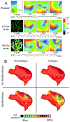
Modeling fibroblast proliferation in the regions of fibrosis. A: Effect of myocyte- fibroblast coupling on spiral wave behavior in a myocardial sheet of size 4.5×4.5 cm. Top, control case without fibroblasts. Middle and bottom, models of low-density and high-density fibroblast proliferation (LD-Fbs and HD-Fbs) in a central circular region of the sheet. In the LD-Fbs and HD-Fbs models, atrial myocytes (100pF), each connecting to 4 fibroblasts (6.3pF) within the Fb-Area, account for 12.5% and 50.0% of that area, respectively. The simulated ECG in each case is shown at the bottom. With permission from. B: Maps of APD in four human atrial models (same atrial geometry). Fibrotic lesions are modeled with (bottom row) and without (top row) myofibroblast infiltration (and coupling to myocytes), as well as with (right column) and without (left column) diffuse collagen deposition for both sets of maps. All models include gap-junction remodeling in the fibrotic lesions. With permission from.
Figure 8
Figure 8
Simulations of AF management. A: Dual stage septal pacing algorithm with successful atrial fibrillation termination in a 3D surface model of the human atria (with permission from). B: Modeling lines of ablation in the atria. Tissue targeted by ablation is shown in white. Modified with permission from.

References

    1. Feinberg WM, Blackshear JL, Laupacis A, Kronmal R, Hart RG. Prevalence, age distribution, and gender of patients with atrial fibrillation. Analysis and implications. Archives of internal medicine. 1995;155:469–473. - PubMed
    1. Miyasaka Y, Barnes ME, Gersh BJ, Cha SS, Bailey KR, Abhayaratna WP, Seward JB, Tsang TS. Secular trends in incidence of atrial fibrillation in olmsted county, minnesota, 1980 to 2000, and implications on the projections for future prevalence. Circulation. 2006;114:119–125. - PubMed
    1. Iwasaki YK, Nishida K, Kato T, Nattel S. Atrial fibrillation pathophysiology: Implications for management. Circulation. 2011;124:2264–2274. - PubMed
    1. Nattel S, Burstein B, Dobrev D. Atrial remodeling and atrial fibrillation: Mechanisms and implications. Circulation. Arrhythmia and electrophysiology. 2008;1:62–73. - PubMed
    1. Schotten U, Verheule S, Kirchhof P, Goette A. Pathophysiological mechanisms of atrial fibrillation: A translational appraisal. Physiological reviews. 2011;91:265–325. - PubMed

Publication types