. 2014 Apr 25;344(6182):380-6.
doi: 10.1126/science.1249656.

Genome sequence of the tsetse fly (Glossina morsitans): vector of African trypanosomiasis

Collaborators

Genome sequence of the tsetse fly (Glossina morsitans): vector of African trypanosomiasis

International Glossina Genome Initiative. Science. .

Abstract

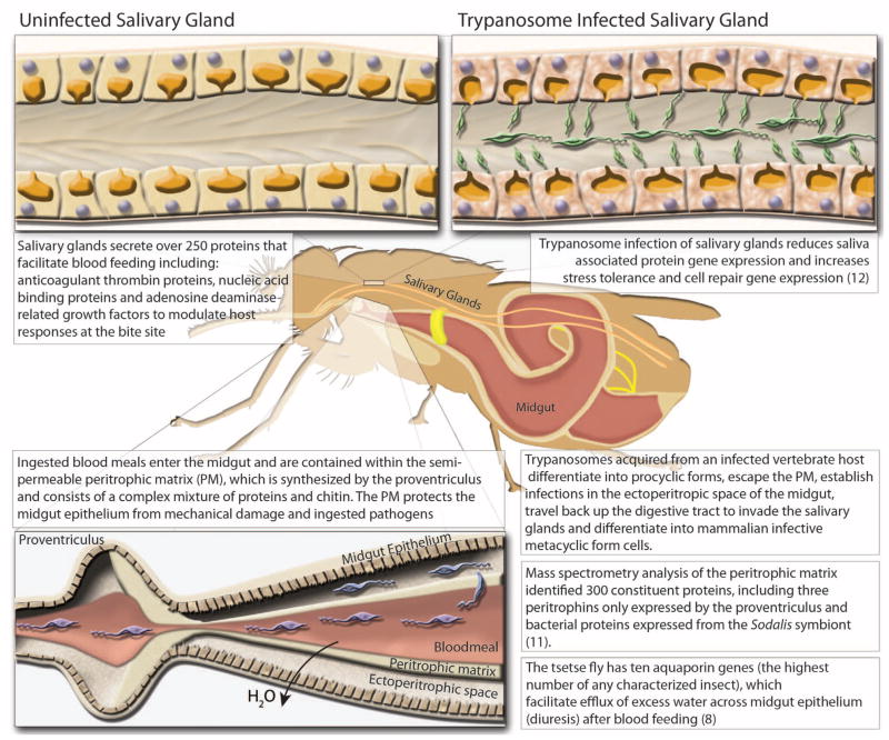
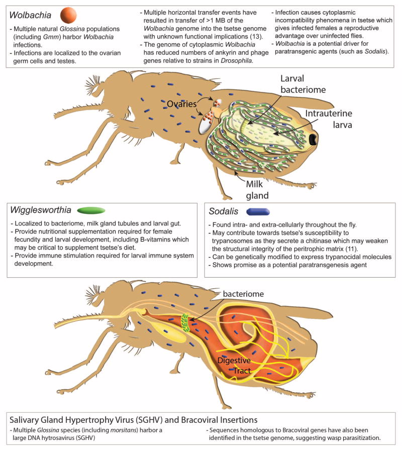
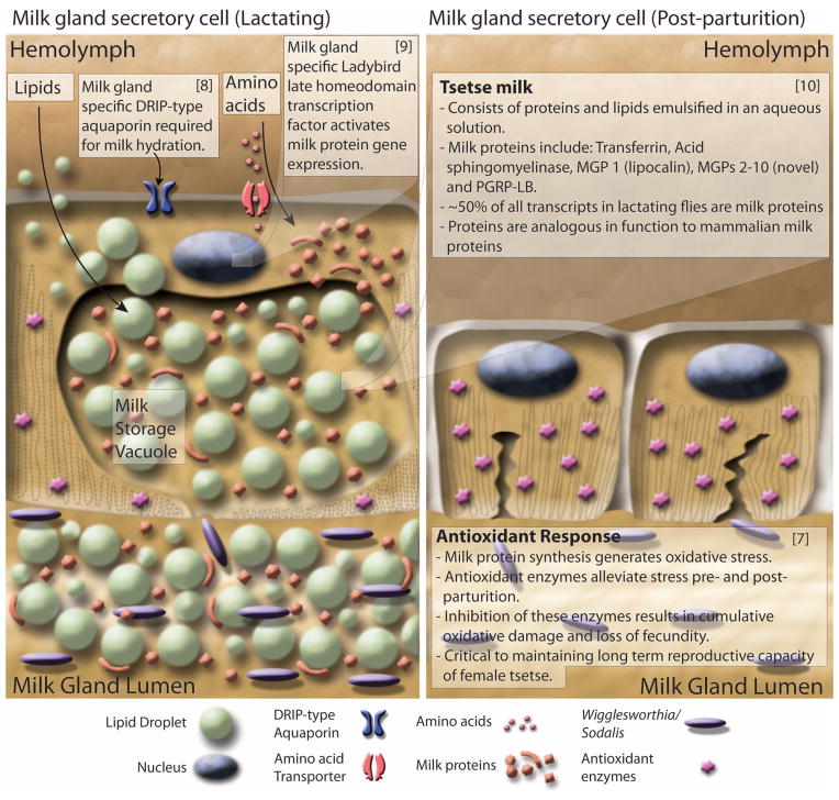
Tsetse flies are the sole vectors of human African trypanosomiasis throughout sub-Saharan Africa. Both sexes of adult tsetse feed exclusively on blood and contribute to disease transmission. Notable differences between tsetse and other disease vectors include obligate microbial symbioses, viviparous reproduction, and lactation. Here, we describe the sequence and annotation of the 366-megabase Glossina morsitans morsitans genome. Analysis of the genome and the 12,308 predicted protein-encoding genes led to multiple discoveries, including chromosomal integrations of bacterial (Wolbachia) genome sequences, a family of lactation-specific proteins, reduced complement of host pathogen recognition proteins, and reduced olfaction/chemosensory associated genes. These genome data provide a foundation for research into trypanosomiasis prevention and yield important insights with broad implications for multiple aspects of tsetse biology.


Figures

Fig. 1
Fig. 1. Diagrammatic presentation of major findings regarding the effects of trypanosome infection of the salivary glands and midgut, proteomic analysis of the peritrophic matrix, and the role of aquaporin proteins in blood meal digestion and diuresis
(Top) comparison of trypanosome-uninfected and -infected states of Glossina salivary glands. (Left) Representative protein components of Glossina salivary secretions. (Right) Pathogenic effects of trypanosome infection on salivary gland function. (Bottom) Glossina digestive physiology and the infection process by trypanosomes. Associated satellite references for these findings are listed within the figure as numbers in parentheses and correspond to the reference list (8, 11, 12).
Fig. 2
Fig. 2. Diagrammatic presentation of the Glossina microbiome, tissue localization of bacterial symbionts, physiological importance, and summary of genomic interactions
(Top) Glossina reproductive physiology and associated symbiont localizations. (Bottom) Glossina digestive physiology and the associated symbiont localizations. Associated text describes significant findings regarding the Glossina microbiome and the associated impacts in terms of Glossina immunity, nutrition, vectorial capacity, and vector control. Associated satellite references are listed within the figure as numbers in parentheses and correspond to the reference list (11, 13).
Fig. 3
Fig. 3. Diagrammatic presentation of milk gland secretory cell physiology and milk production during lactation and after parturition
(Left) Lactation. Nutrients including lipids, amino acids, and water are taken up by the cell through various transporters. Lipids are aggregated into droplets while amino acids are incorporated into the synthesis of a battery of milk proteins. During pregnancy, milk protein genes are up-regulated by the Ladybird Late homeodomain protein. Lipids, proteins, and water are combined to form the milk constituents, which are stored in a large extracellular secretory reservoir. Stored milk is secreted into the lumen, which also houses the extracellular obligate bacterial symbiont Wigglesworthia. The milk and symbionts are transported through the gland to the uterus, where the developing larvae feeds upon these secretions. (Right) Involution and recovery. After parturition, milk gland cells shrink, undergo autophagy, and express antioxidant enzymes to inhibit oxidative damage. The recovered cells prepare for the next round of lactation by regeneration of protein synthesis and structural components. Associated satellite references for these findings are listed within the figure as numbers in parentheses and correspond to the reference list (–10).

Comment in

Similar articles

Cited by

References

    1. Welburn SC, Maudlin I, Simarro PP. Parasitology. 2009;136:1943–1949. - PubMed
    1. Wiegmann BM, et al. Proc Natl Acad Sci USA. 2011;108:5690–5695. - PMC - PubMed
    1. Petersen FT, Meier R, Kutty SN, Wiegmann BM. Mol Phylogenet Evol. 2007;45:111–122. - PubMed
    1. Lehane MJ, Aksoy S, Levashina E. Trends Parasitol. 2004;20:433–439. - PubMed
    1. Weiss BL, Wang J, Maltz MA, Wu Y, Aksoy S. PLOS Pathog. 2013;9:e1003318. - PMC - PubMed

Publication types

MeSH terms

Substances

Associated data