. 2014 Jul 1;20(13):3472-84.
doi: 10.1158/1078-0432.CCR-13-2944. Epub 2014 Apr 24.

Regorafenib inhibits colorectal tumor growth through PUMA-mediated apoptosis

Affiliations

Regorafenib inhibits colorectal tumor growth through PUMA-mediated apoptosis

Dongshi Chen et al. Clin Cancer Res. .

Abstract

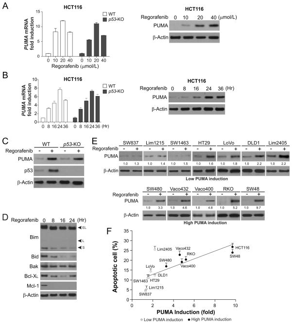
Purpose: Regorafenib, a multikinase inhibitor targeting the Ras/Raf/MEK/ERK pathway, has recently been approved for the treatment of metastatic colorectal cancer. However, the mechanisms of action of regorafenib in colorectal cancer cells have been unclear. We investigated how regorafenib suppresses colorectal cancer cell growth and potentiates effects of other chemotherapeutic drugs.

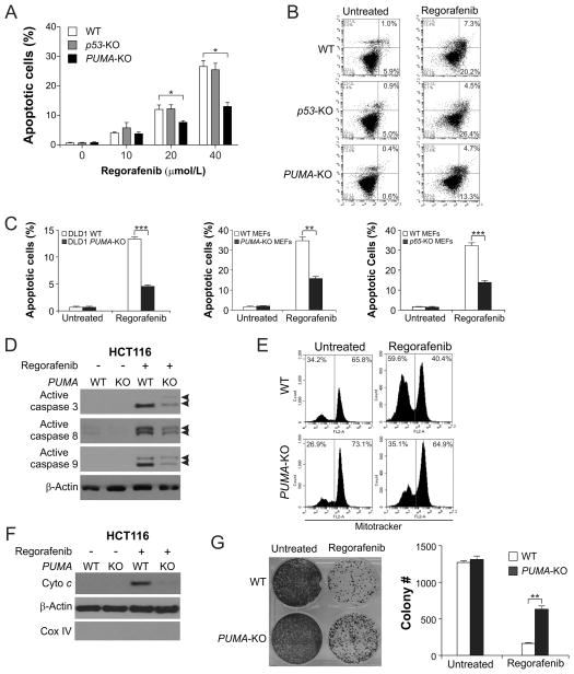
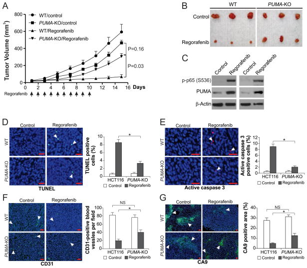
Experimental design: We determined whether and how regorafenib induces the expression of PUMA, a p53 target and a critical mediator of apoptosis in colorectal cancer cells. We also investigated whether PUMA is necessary for the killing and chemosensitization effects of regorafenib in colorectal cancer cells. Furthermore, xenograft tumors were used to test if PUMA mediates the in vivo antitumor, antiangiogenic, and chemosensitization effects of regorafenib.

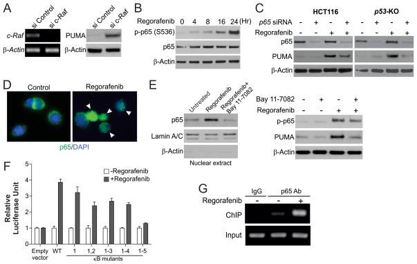
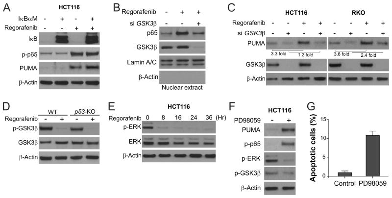
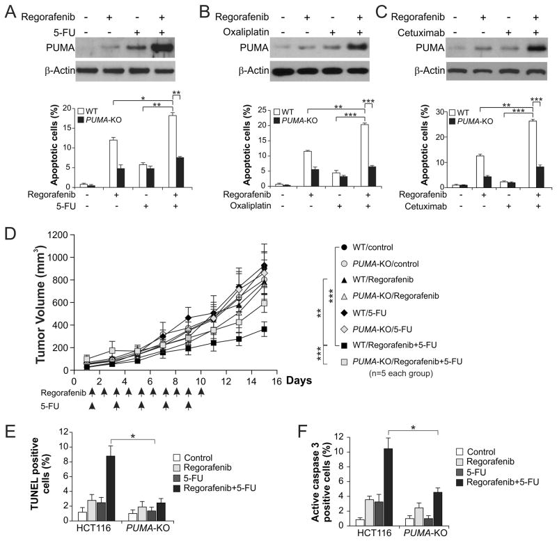
Results: We found that regorafenib treatment induces PUMA in colorectal cancer cells irrespective of p53 status through the NF-κB pathway following ERK inhibition and glycogen synthase kinase 3β activation. Upregulation of PUMA is correlated with apoptosis induction in different colorectal cancer cell lines. PUMA is necessary for regorafenib-induced apoptosis in colorectal cancer cells. Chemosensitization by regorafenib is mediated by enhanced PUMA induction through different pathways. Furthermore, deficiency in PUMA abrogates the in vivo antitumor, antiangiogenic, and chemosensitization effects of regorafenib.

Conclusions: Our results demonstrate a key role of PUMA in mediating the anticancer effects of regorafenib in colorectal cancer cells. They suggest that PUMA induction can be used as an indicator of regorafenib sensitivity, and also provide a rationale for manipulating the apoptotic machinery to improve the therapeutic efficacy of regorafenib and other targeted drugs.


Conflict of interest statement

Disclosures: The authors declare no conflict of interest and all authors have agreed on the submission.

Figures

Figure 1
Figure 1. Upregulation of PUMA expression by regorafenib correlates with apoptosis induction in CRC cells
(A) WT and p53-knockout (p53-KO) HCT116 colon cancer cells were treated with regorafenib at indicated concentrations for 24 hours. Left, PUMA mRNA induction by regorafenib was analyzed by real-time reverse transcriptase (RT) PCR, with β-actin as a control. Right, PUMA and β-actin expression was analyzed by Western blotting. (B) WT and p53-KO HCT116 cells were treated with 40 μmol/L regorafenib and analyzed at different time points after treatment. Left, time course of PUMA mRNA induction was determined by real-time RT PCR, with β-actin as a control. Right, time course of PUMA protein induction was analyzed by Western blotting. (C) WT and p53-KO HCT116 cells were treated with 40 μmol/L regorafenib for 24 hours. PUMA expression was analyzed by Western blotting. (D) The expression of indicated Bcl-2 family members was analyzed by Western blotting in HCT116 cells treated with 40 μmol/L regorafenib at indicated time points. (E) Western blot analysis of PUMA expression in indicated CRC cell lines treated with 40 μmol/L regorafenib for 24 hours. Relative PUMA expression, which was quantified by Image J program and normalized to that of β-actin, is indicated, with that in untreated cells arbitrarily set as 1.0. (F) Indicated CRC cell lines were treated with 40 μmol/L regorafenib for 48 hours. Apoptosis was quantified by counting condensed and fragmented nuclei after nuclear staining with Hoechst 33258, and plotted against PUMA induction from (E). The results represent means + SD of 3 independent experiments.
Figure 2
Figure 2. PUMA mediates the apoptotic and anticancer effects of regorafenib through the mitochondrial pathway
(A) WT, p53-KO, and PUMA-KO HCT116 cells were treated with regorafenib at indicated concentrations for 48 hours. Apoptosis was analyzed by counting condensed and fragmented nuclei after nuclear staining with Hoechst 33258. (B) Apoptosis in cells treated with 40 μmol/L regorafenib for 48 hours was analyzed by annexin V/PI staining followed by flow cytometry. The percentages of annexin-positive apoptotic cells are indicated in the two right quadrants. (C) Comparison of apoptosis in WT and PUMA-KO DLD1 colon cancer cells (left), WT and PUMA-KO MEFs (middle), and WT and p65-KO MEFs (right) following treatment with 40 μmol/L regorafenib for 48 hours. Apoptosis was analyzed by nuclear staining as in (A). (D) Western blot analysis of active caspase 3, active caspase 8, and active caspase 9 (indicated by arrow heads) in WT and PUMA-KO HCT116 cells with or without regorafenib (40 μmol/L) treatment for 24 hours. (E) After treatment of WT and PUMA-KO HCT116 cells with 40 μmol/L regorafenib for 36 hours, mitochondrial membrane potential was analyzed by flow cytometry following MitoTracker Red CMXRos staining. (F) Cytosolic fractions isolated from WT and PUMA-KO HCT116 cells treated with 40 μmol/L regorafenib for 36 hours were probed for cytochrome c by Western blotting. α-Tubulin and cytochrome oxidase subunit IV (Cox IV), which are expressed in cytoplasm and mitochondria, respectively, were analyzed as the control for loading and fractionation. (G) Colony formation of WT and PUMA-KO HCT116 cells treated with 40 μmol/L regorafenib for 48 hours at 14 days following crystal violet staining of attached cells. Left, representative pictures of colonies; Right, quantification of colony numbers. Results in (A), (C), and (G) were expressed as means ± SD of 3 independent experiments. ***, P <0.001; **, P <0.01; *, P <0.05.
Figure 3
Figure 3. Activation of p65 mediates PUMA induction in response to regorafenib treatment
(A) HCT116 cells were transfected with either a control scrambled siRNA or a c-Raf siRNA for 24 hours, and then treated with 40 μmol/L regorafenib for 24 hours. c-Raf mRNA and PUMA protein expression were analyzed by RT-PCR and Western blotting, respectively. (B) HCT116 cells were treated with 40 μmol/L regorafenib. Expression of p-p65 (S536) and β-actin at indicated time points was analyzed by Western blotting. (C) WT and p53-KO HCT116 cells were transfected with either a control scrambled siRNA or a p65 siRNA for 24 hours, and then treated with 40 μmol/L regorafenib for 24 hours. p65 and PUMA expression was analyzed by Western blotting. (D) HCT116 cells were treated with 40 μmol/L regorafenib for 3 hours and then fixed. Immunofluorescence was carried out as described in the Materials and Methods for p65 (green) and DAPI (blue). Representative pictures (400×) are shown. Arrows indicate cells with p65 nuclear translocation. (E) HCT116 cells were treated with 10 μmol/L BAY11-7082 for 1 hour, and then with 40 μmol/L regorafenib for 24 hours. Left, nuclear fractions were isolated from cells and analyzed for p65 expression by Western blotting. Lamin A/C and β-actin, which are expressed in nucleus and cytoplasm, respectively, were used as controls for loading and fractionation. Right, the levels of p-p65 (S536) and PUMA were analyzed by Western blotting. (F) p53-KO HCT116 cells were transfected overnight with a PUMA promoter luciferase reporter containing WT κB sites or indicated mutants, and then treated with 40 μmol/L regorafenib for 16 hours. Reporter activities were normalized to the untreated control samples and plotted. (G) Chromatin immunoprecipitation (ChIP) was performed using anti-p65 antibody on HCT116 cells following regorafenib (40 μmol/L) treatment for 8 hours. The IgG was used to control for antibody specificity. PCR was carried out using primers surrounding the p65 binding sites in the PUMA promoter.
Figure 4
Figure 4. Regorafenib activates GSK3β and inhibits ERK to induce PUMA through p65
(A) HCT116 cells were transfected with pCMV or IκBαM overnight, and then treated with 40 μmol/L regorafenib for 24 hours. PUMA, p-p65 (S536) and IκB were analyzed by Western blotting. (B) HCT116 cells were transfected with either a control scrambled siRNA or a GSK3β siRNA for 24 hours, and then treated with 40 μmol/L regorafenib for 3 hours. Nuclear fractions were isolated from cells treated with regorafenib and analyzed for p65 and GSK3β expression by Western blotting. (C) After GSK3β siRNA transfection as in (B), HCT116 and RKO cells were treated with 40 μmol/L regorafenib for 24 hours. GSK3β and PUMA were analyzed by Western blotting. PUMA induction was determined by quantifying PUMA expression using Image J program and normalizing to that of β-actin. (D) The levels of total GSK3β and p-GSK3β (S9) were analyzed by Western blotting in WT and p53-KO HCT116 cells treated with 40 μmol/L regorafenib for 24 hours. (E) The levels of total ERK and p-ERK (T202/Y204) were analyzed by Western blotting in HCT116 cells treated with 40 μmol/L regorafenib at indicated time points. (F) HCT116 cells were treated with 25 μmol/L of the ERK inhibitor PD98059 for 24 hours. The levels of PUMA, p-GSK3β (S9), p-ERK1/2 (T202/Y204) and p-p65 (S536) were analyzed by Western blotting. (G) Apoptosis in HCT116 cells treated with PD98059 as in (F) for 48 hours was determined by nuclear staining with Hoechst 33258.
Figure 5
Figure 5. The antitumor effects of regorafenib in vivo are PUMA-dependent
(A) Nude mice were injected s.c. with 4 × 10⁶ WT or PUMA-KO HCT116 cells. After 1 week, mice were oral gavaged with 30 mg/kg regorafenib or the vehicle control cremephor EL/ethanol for 10 consecutive days. Tumor volume at indicated time points after treatment was calculated and plotted with p values, n=5 in each group. Arrows indicate regorafenib injection. (B) Representative tumors at the end of the experiment in (A). (C) Mice with WT HCT116 xenograft tumors were treated with 30 mg/kg regorafenib or the vehicle as in (A) for 4 consecutive days. The levels of p-p65 (S536) and PUMA in randomly selected tumors were analyzed by Western blotting. (D) Paraffin-embedded sections of WT or PUMA-KO tumor tissues from mice treated as in (C) were analyzed by TUNEL staining. Left, representative TUNEL staining pictures; Right, TUNEL-positive cells were counted and plotted. (E) Tissue sections from (D) were analyzed by active caspase 3 staining. Left, representative staining pictures; Right, active caspase 3-positive cells were counted and plotted. (F) Tissue sections from (B) were analyzed for blood vessel formation by CD31 staining. Left, representative staining pictures; Right, CD31-positive cells were counted and plotted. (G) Tissue sections from (B) were analyzed for hypoxia by CA9 staining. Left, representative staining pictures; Right, CA9-positive areas were quantified by Image J program and plotted. In (D)–(G), results were expressed as means ± SD of 3 independent experiments. Arrows, example cells with positive staining; scale bars, 25 μm; *, P <0.05; NS, P > 0.05.
Figure 6
Figure 6. PUMA mediates the chemosensitization effects of regorafenib in vitro and in vivo
(A) WT and PUMA-KO HCT116 cells were treated with 20 μmol/L regorafenib, 20 mg/L 5-fluorouracil (5-FU), or their combination. Upper, western blot analysis of PUMA expression in WT cells treated for 24 hours; lower, apoptosis in WT and PUMA-KO cells treated for 48 hours analyzed by nuclear staining with Hoechst 33258. (B) WT and PUMA-KO HCT116 cells were treated with 20 μmol/L regorafenib, 25 μmol/L oxaliplatin, or their combination. Upper, western blot analysis of PUMA expression in WT cells treated for 24 hours; lower, apoptosis in WT and PUMA-KO cells treated for 48 hours analyzed as in (A). (C) WT and PUMA-KO HCT116 cells were treated with 20 μmol/L regorafenib, 6 μg/mL of cetuximab, or their combination. Upper, western blot analysis of PUMA expression in WT cells treated for 24 hours; lower, apoptosis in WT and PUMA-KO cells treated for 48 hours analyzed as in (A). (D) Nude mice were injected s.c. with 4 × 10⁶ WT or PUMA-KO HCT116 cells. After 1 week, mice were treated with 15 mg/kg regorafenib daily by oral gavage, 25 mg/kg 5-FU every other day by i.p. injection, or their combination for 10 consecutive days. Tumor volume at indicated time points after treatment was calculated and plotted with p values for indicated comparisons, n=5 in each group. Arrows indicate regorafenib or 5-FU injection. (E) Mice with WT or PUMA-KO HCT116 xenograft tumors were treated with 15 mg/kg regorafenib daily, 25 mg/kg 5-FU every other day, or their combination as in (D) for 4 consecutive days. Paraffin-embedded sections were analyzed by TUNEL staining. TUNEL-positive cells were counted and plotted. (F) Tissue sections from (E) were analyzed by active caspase 3 staining. Active caspase 3-positive cells were counted and plotted. Results in (E) and (F) were expressed as means ± SD of 3 independent experiments. ***, P <0.001; **, P <0.01; *, P <0.05.

References

    1. Siegel R, Naishadham D, Jemal A. Cancer statistics, 2012. CA Cancer J Clin. 2012;62:10–29. - PubMed
    1. Poston GJ, Figueras J, Giuliante F, Nuzzo G, Sobrero AF, Gigot JF, et al. Urgent need for a new staging system in advanced colorectal cancer. J Clin Oncol. 2008;26:4828–33. - PubMed
    1. Segal NH, Saltz LB. Evolving treatment of advanced colon cancer. Annual review of medicine. 2009;60:207–19. - PubMed
    1. Chu E. An update on the current and emerging targeted agents in metastatic colorectal cancer. Clinical colorectal cancer. 2012;11:1–13. - PubMed
    1. Martini M, Vecchione L, Siena S, Tejpar S, Bardelli A. Targeted therapies: how personal should we go? Nat Rev Clin Oncol. 2012;9:87–97. - PubMed

Publication types

MeSH terms