. 2014 Jun 6;289(23):15904-14.
doi: 10.1074/jbc.M114.555631. Epub 2014 Apr 24.

Vipp1 is essential for the biogenesis of Photosystem I but not thylakoid membranes in Synechococcus sp. PCC 7002

Affiliations

Vipp1 is essential for the biogenesis of Photosystem I but not thylakoid membranes in Synechococcus sp. PCC 7002

Shuyi Zhang et al. J Biol Chem. .

Abstract

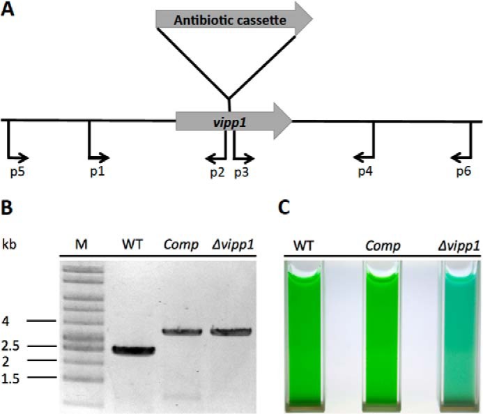
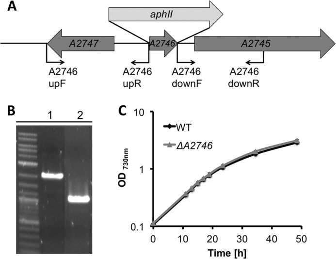
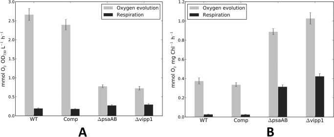
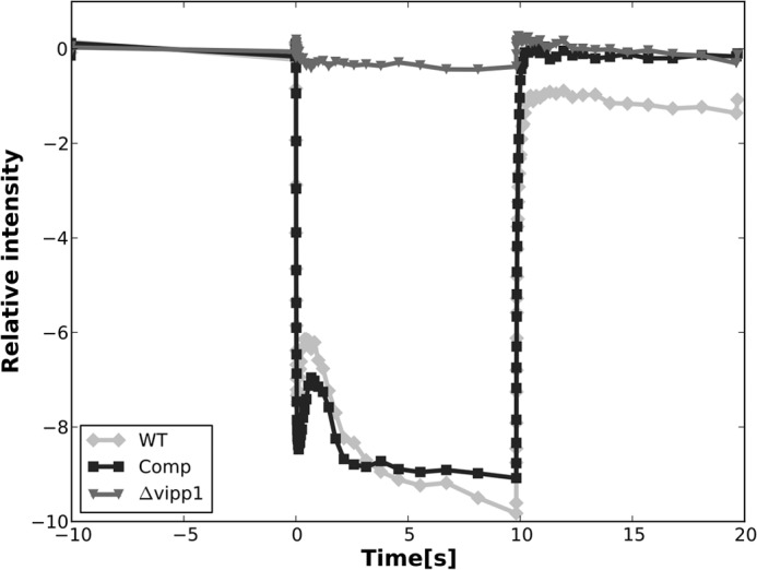
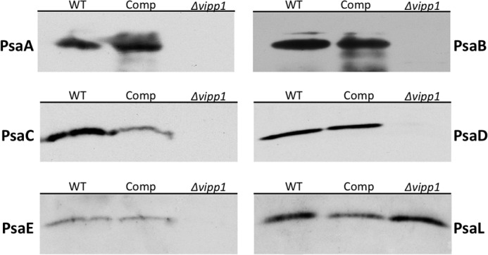
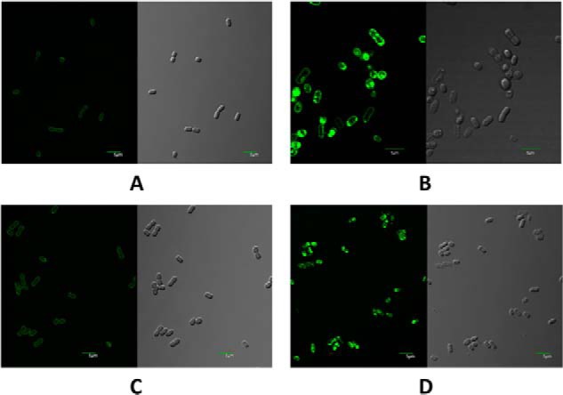
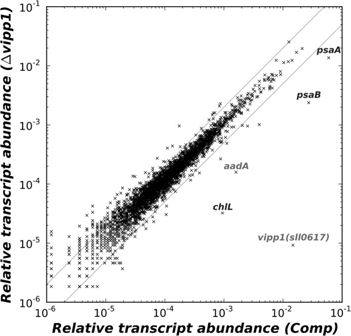
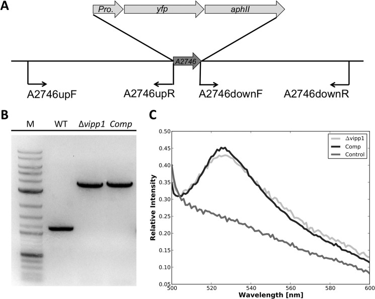
The biogenesis of thylakoid membranes in cyanobacteria is presently not well understood, but the vipp1 gene product has been suggested to play an important role in this process. Previous studies in Synechocystis sp. PCC 6803 reported that vipp1 (sll0617) was essential. By constructing a fully segregated null mutant in vipp1 (SynPCC7002_A0294) in Synechococcus sp. PCC 7002, we show that Vipp1 is not essential. Spectroscopic studies revealed that Photosystem I (PS I) was below detection limits in the vipp1 mutant, but Photosystem II (PS II) was still assembled and was active. Thylakoid membranes were still observed in vipp1 mutant cells and resembled those in a psaAB mutant that completely lacks PS I. When the vipp1 mutation was complemented with the orthologous vipp1 gene from Synechocystis sp. PCC 6803 that was expressed from the strong P(cpcBA) promoter, PS I content and activities were restored to normal levels, and cells again produced thylakoids that were indistinguishable from those of wild type. Transcription profiling showed that psaAB transcripts were lower in abundance in the vipp1 mutant. However, when the yfp gene was expressed from the P(psaAB) promoter in the presence and the absence of Vipp1, no difference in YFP expression was observed, which shows that Vipp1 is not a transcription factor for the psaAB genes. This study shows that thylakoids are still produced in the absence of Vipp1 and that normal thylakoid biogenesis in Synechococcus sp. PCC 7002 requires expression and biogenesis of PS I, which in turn requires Vipp1.

Keywords: Cyanobacteria; Electron Microscopy (EM); Membrane Biogenesis; Photosynthesis; Photosystem I; Protein Translocation.


Figures

FIGURE 1.
FIGURE 1.
Construction and verification of vipp1 mutant as well as a trans-complemented strain. A, scheme showing the construction of a vipp1 mutant by homologous recombination by using primer set p1 and p2 to amplify the upstream region and primer set p3 and p4 to amplify the downstream region. An antibiotic resistance cassette was ligated into restriction sites added to the appropriate ends of the flanking sequences (see “Experimental Procedures” for other details). B, results of agarose gel electrophoresis of amplicons produced using primers p5 and p6, showing the fully segregated interruption mutant of vipp1. The template DNAs were isolated from the wild type (WT), the trans-complemented vipp1 mutant (Comp), and the vipp1 mutant strain (Δvipp1). The results clearly showed that the wild-type vipp1 and interrupted vipp1::aacC1 alleles had segregated completely in the vipp1 mutant and that the vipp1 gene was still mutated in the Comp strain. Lane M, DNA size markers. C, cultures of equivalent cell density (OD730 nm = 1.0) for WT, Comp, and Δvipp1.
FIGURE 2.
FIGURE 2.
Construction and verification of a neutral site platform for Synechococcus sp. PCC 7002. A, diagram showing the construction of a SynPCC7002_A2746 mutant and positions of oligonucleotide primers (see Table 1). B, agarose gel electrophoresis of amplicons produced by polymerase chain reaction, demonstrating complete segregation of alleles for SynPCC7002_A2746::aphII and SynPCC7002_A2746, using primer set A2746upF and A2746downR. The template DNAs were isolated from the SynPCC7002_A2746 mutant (lane 1) and wild type (lane 2). C, comparison of growth rates for Synechococcus sp. PCC 7002 WT and a neutral site mutant strain constructed in open reading frame SynPCC7002_A2746 (ΔA2746). The growth rates were indistinguishable within experimental error. The data are the average of three biological replicates.
FIGURE 3.
FIGURE 3.
Immunoblotting of whole cell extracts of Synechococcus sp. PCC 7002 strains with antibodies to Vipp1. Antibodies to Vipp1 were used to detect Vipp1 levels in WT, Comp, Δvipp1, and a PS I-less mutant (ΔpsaAB). Equal quantities of cells were used to produce the extracts for this experiment, so the Vipp1 levels detected in this experiment can be compared semiquantitatively.
FIGURE 4.
FIGURE 4.
Thylakoid membrane morphology as revealed by electron microscopy. Thin sections of Synechococcus sp. PCC 7002 cells from various strains were examined by transmission electron microscopy. A, thylakoid membranes were normally assembled in WT (A) and in Comp (C). Thylakoids were greatly reduced in number and area in the vipp1 mutant (B), and these cells closely resembled cells for a psaAB mutant (D). The arrows in the bottom right of B show thylakoids that clearly appear to be directly connected to the cytoplasmic membrane. Bars, 500 nm.
FIGURE 5.
FIGURE 5.
Oxygen evolution and respiration rates for WT, Comp, ΔpsaAB, and Δvipp1, based on equal cell numbers (based on OD730 nm) (A) or equal Chl a (B). The vipp1 mutant strain as well as the psaAB mutant strain had much higher oxygen evolution rates than the WT strain when rates were compared on the basis of Chl but much lower oxygen evolution rates when rates were compared on the basis of equal cell numbers. Note that these values were derived from the initial rates of oxygen evolution for the vipp1 and the psaAB mutant strains, because oxygen evolution rates rapidly declined for these two strains that had no PS I activity to drive the reoxidation of the plastoquinone pool. The data shown are averages values for three biological replicates, and the error bars show the standard deviation.
FIGURE 6.
FIGURE 6.
Low temperature (77 K) fluorescence emission spectra of whole cells of WT, Comp, and Δvipp1. In the Δvipp1 mutant, PS I fluorescence emission at ∼715 nm was completely absent, but PS II was still assembled and exhibited normal fluorescence emission at 685 and 695 nm. The excitation wavelength was 440 nm.
FIGURE 7.
FIGURE 7.
Photobleaching of P700 in whole cells of WT (light gray line), Comp (black line), and Δvipp1 (dark gray line). The PS I activity in the trans-complemented vipp1 mutant strain was almost the same as that of the WT. No P700 photobleaching activity was detected in the vipp1 mutant strain. The actinic light was turned on at 0 s and turned off after 10 s, and absorption difference was measured at 700 nm.
FIGURE 8.
FIGURE 8.
Immunoblotting of PS I subunits. Antibodies to PsaA, PsaB, PsaC, PsaD, PsaE, and PsaL were used to detect the presence of the PS I subunits in whole cell extracts of WT, Comp, and Δvipp1. PsaL was the only PS I subunit detected in the vipp1 mutant strain, but all subunits were detected in the WT control and Comp strains. Note that these immunoblots were performed with whole cell extracts of cells scraped from plates; thus, the results are only qualitative and should not be compared quantitatively.
FIGURE 9.
FIGURE 9.
Confocal fluorescence microscopy images (left) and differential interference contrast images (right) of Synechococcus sp. PCC 7002 cells stained with Nile red. For the fluorescence images, the excitation wavelength was 488 nm, and emitted light in the wavelength range from 500 to 600 nm was detected. Scale bars, 5 μm. A, cells of wild-type Synechococcus sp. PCC 7002. B, cells of the vipp1 mutant strain. C, trans-complemented cells of the vipp1 mutant strain. D, cells of the psaAB mutant strain.
FIGURE 10.
FIGURE 10.
Scatter plot comparing the relative transcript abundances for mRNAs of the vipp1 mutant to those in the trans-complemented vipp1 mutant strain. Transcripts for psaA and psaB were specifically higher in the trans-complemented vipp1 strain, which also had enhanced mRNA levels for the vipp1 (sll0617) gene from Synechocystis sp. PCC 6803. Transcript levels for chlL were also more abundant in the trans-complemented strain, but nearly all other transcripts were unchanged. The gray lines indicate 2-fold changes in transcript levels.
FIGURE 11.
FIGURE 11.
Construction (A) and verification (B) of insertion of PpsaAB or PchlLN promoters fused to yfp into a neutral site in the chromosome of Synechococcus sp. PCC 7002 strains and relative YFP fluorescence emission in the resulting strains (C). A, scheme showing the replacement of open reading frame SynPCC7002_A2746 by yfp reporter gene fusions by homologous recombination and selection with kanamycin (aphII gene cassette confers resistance to kanamycin). Promoter regions (Pro.) for psaAB or chlLN were transcriptionally fused to the yfp gene, which was placed upstream from an aphII gene, which encodes aminoglycoside phosphotransferase II and confers resistance to kanamycin. B, agarose gel electrophoresis of PCR amplicons using primer set A2746upF and A2746downR to verify the introduction of the promoter-yfp-aphII constructions in the respective strains. Template DNAs came from the WT strain, the vipp1 mutant, and the Comp. C, fluorescence emission spectra showing YFP emission in the constructed reporter strains. The control spectrum shows fluorescence emission from the vipp1 strain, which does not contain the yfp gene. Very similar levels of YFP fluorescence emission were detected in the vipp1 and Comp strains, which carried the yfp gene fused to the PpsaAB promoter. These data indicate that Vipp1 does not modify transcription from the PpsaAB promoter.

Similar articles

Cited by

References

    1. De Marais D. J. (2000) When did photosynthesis emerge on Earth? Science 289, 1703–1705 - PubMed
    1. Jordan P., Fromme P., Witt H. T., Klukas O., Saenger W., Krauss N. (2001) Three-dimensional structure of cyanobacterial photosystem I at 2.5 Å resolution. Nature 411, 909–917 - PubMed
    1. Vothknecht U. C., Westhoff P. (2001) Biogenesis and origin of thylakoid membranes. Biochim. Biophys. Acta 1541, 91–101 - PubMed
    1. Kroll D., Meierhoff K., Bechtold N., Kinoshita M., Westphal S., Vothknecht U. C., Soll J., Westhoff P. (2001) VIPP1, a nuclear gene of Arabidopsis thaliana essential for thylakoid membrane formation. Proc. Natl. Acad. Sci. U.S.A. 98, 4238–4242 - PMC - PubMed
    1. Li H.-M., Kaneko Y., Keegstra K. (1994) Molecular cloning of a chloroplastic proteinassociated with both the envelope and thylakoid membranes. Plant Mol. Biol. 25, 619–632 - PubMed

Publication types

LinkOut - more resources