. 2014 Jul 3;124(1):13-23.
doi: 10.1182/blood-2014-02-558114. Epub 2014 Apr 24.

Requirement for CDK6 in MLL-rearranged acute myeloid leukemia

Affiliations

Requirement for CDK6 in MLL-rearranged acute myeloid leukemia

Theresa Placke et al. Blood. .

Abstract

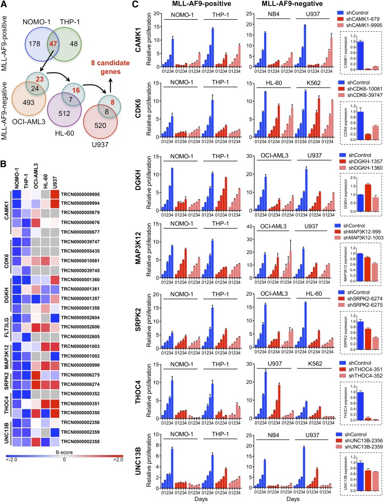
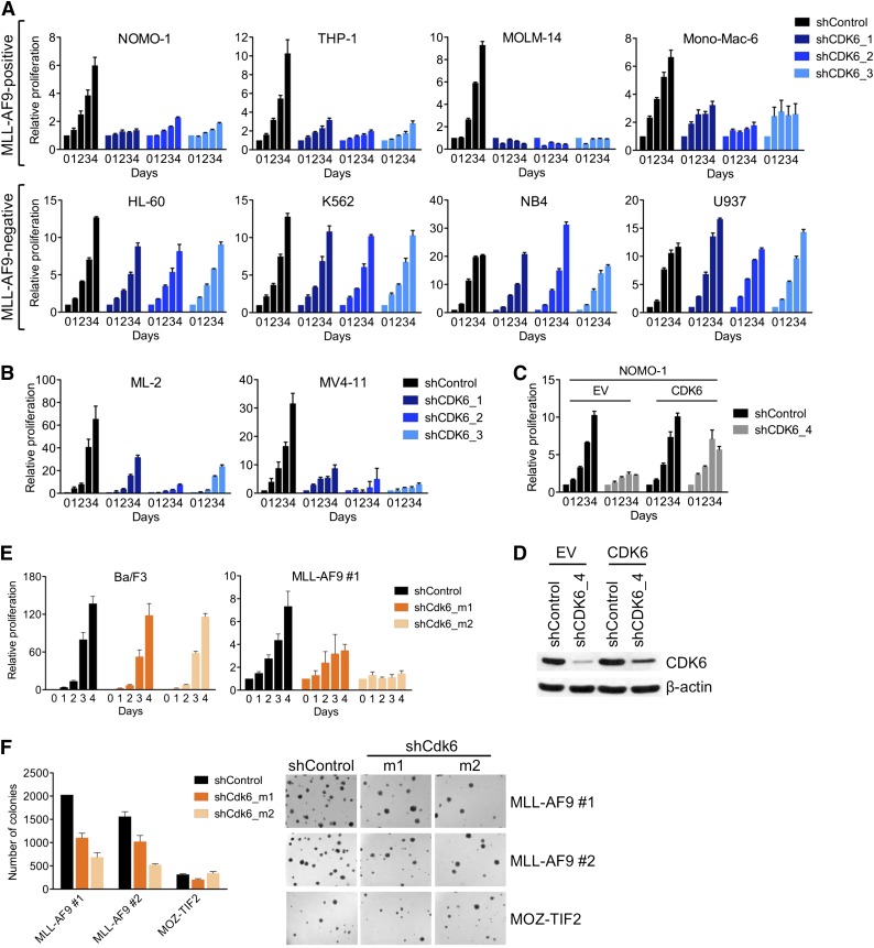
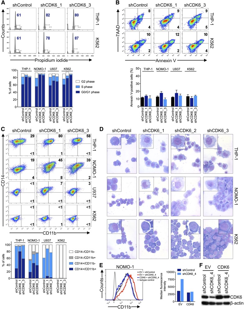
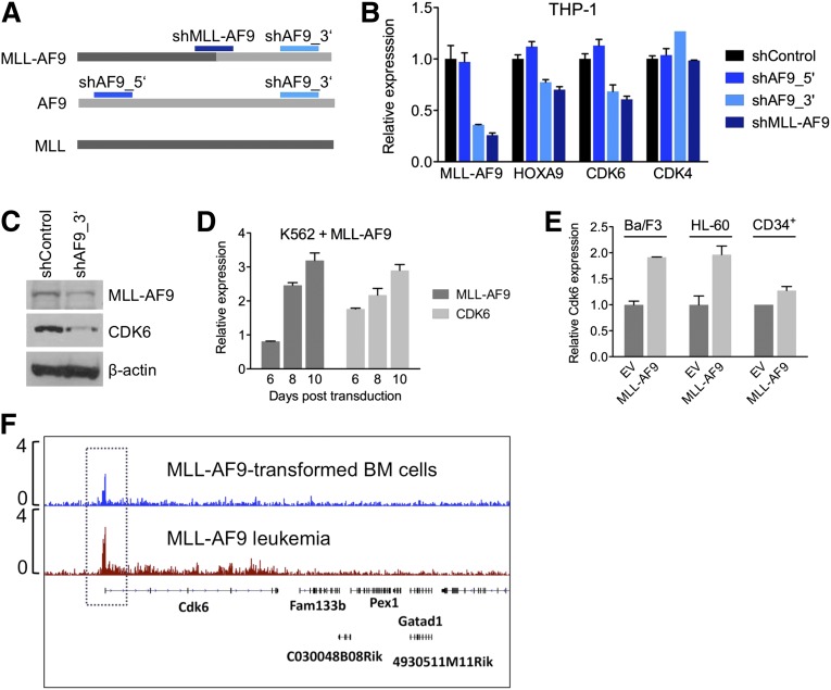
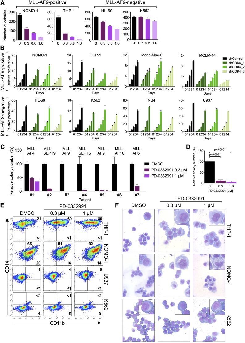
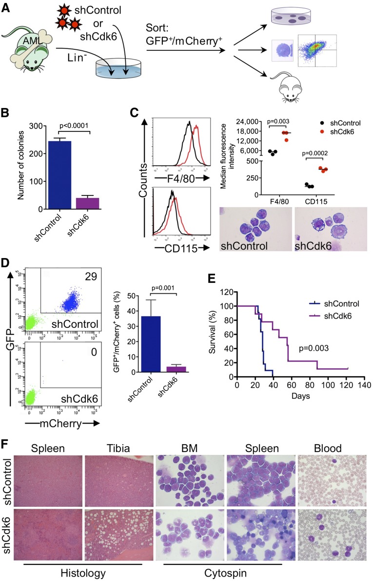
Chromosomal rearrangements involving the H3K4 methyltransferase mixed-lineage leukemia (MLL) trigger aberrant gene expression in hematopoietic progenitors and give rise to an aggressive subtype of acute myeloid leukemia (AML). Insights into MLL fusion-mediated leukemogenesis have not yet translated into better therapies because MLL is difficult to target directly, and the identity of the genes downstream of MLL whose altered transcription mediates leukemic transformation are poorly annotated. We used a functional genetic approach to uncover that AML cells driven by MLL-AF9 are exceptionally reliant on the cell-cycle regulator CDK6, but not its functional homolog CDK4, and that the preferential growth inhibition induced by CDK6 depletion is mediated through enhanced myeloid differentiation. CDK6 essentiality is also evident in AML cells harboring alternate MLL fusions and a mouse model of MLL-AF9-driven leukemia and can be ascribed to transcriptional activation of CDK6 by mutant MLL. Importantly, the context-dependent effects of lowering CDK6 expression are closely phenocopied by a small-molecule CDK6 inhibitor currently in clinical development. These data identify CDK6 as critical effector of MLL fusions in leukemogenesis that might be targeted to overcome the differentiation block associated with MLL-rearranged AML, and underscore that cell-cycle regulators may have distinct, noncanonical, and nonredundant functions in different contexts.


Figures

Figure 1
Figure 1
RNAi screens for genes required by MLL-AF9pos AML cell lines. (A) Schematic of RNAi screens. Numbers in circles indicate genes scoring as positive. Genes scoring exclusively in MLL-AF9pos AML cell lines are indicated in red. (B) Candidate genes preferentially required in MLL-AF9pos AML cell lines. For each of the top-ranking genes, the shRNAs scoring in NOMO-1 and/or THP-1 are shown. Negative B-scores indicate reduced viable cell numbers. (C) Validation of candidate genes. Shown are the effects of 2 shRNAs per candidate gene on cell viability and proliferation of AML cell lines with and without an MLL-AF9 fusion and the knockdown efficiency of each shRNA in NOMO-1 cells, as determined by qRT-PCR.
Figure 2
Figure 2
Requirement for CDK6 in MLL-rearranged hematopoietic cells. (A) Effects of CDK6 suppression in MLL-AF9pos and MLL-AF9neg AML cell lines. (B) Effects of CDK6 suppression in MLL-AF4pos MV4-11 and MLL-AF6pos ML-2 cells. (C) Rescue of viable cell number by expression of the CDK6 coding sequence in NOMO-1 cells transduced with an shRNA targeting the CDK6 3′ UTR. (D) CDK6 protein expression of cells used in panel C. (E) Effects of Cdk6 suppression in suspension cultures of Ba/F3 cells and murine HSPC transduced with MLL-AF9. (F) Effects of Cdk6 suppression in methylcellulose cultures of murine HSPC transduced with MLL-AF9 or MOZ-TIF2. Original magnification, ×25.
Figure 3
Figure 3
Effects of CDK6 suppression in AML cells. (A) Flow cytometric analysis of cell-cycle progression. Numbers indicate percentages of cells in G0/G1. (B) Flow cytometric analysis of apoptosis. Numbers indicate percentages of cells. (C) Flow cytometric analysis of myeloid differentiation in AML cell lines. Numbers indicate percentages of cells. (D) Microscopic analysis of May-Grünwald-Giemsa–stained cytospin preparations of AML cell lines. Original magnification, ×400. Insets show twofold magnified details of the corresponding photographs. (E) Inhibition of myeloid differentiation by expression of the CDK6 coding sequence in NOMO-1 cells transduced with an shRNA targeting the CDK6 3′ UTR. (F) Immunoblot analysis of cells shown in panel E.
Figure 4
Figure 4
Transcriptional activation of CDK6 by MLL-AF9. (A) Schematic of the regions in MLL-AF9 and AF9 targeted by the shRNAs used in panels B and C. (B) Expression of HOXA9, CDK6, and CDK4 mRNA in THP-1 cells after MLL-AF9 suppression. (C) Expression of CDK6 protein in THP-1 cells after MLL-AF9 suppression. (D) Expression over time of MLL-AF9 and CDK6 in K562 cells transduced with MLL-AF9. (E) Expression of CDK6 in HL-60 cells, normal human CD34pos cells, and murine Ba/F3 cells transduced with MLL-AF9. (F) ChIP-seq analysis identifying Cdk6 as direct MLL-AF9 target gene in mouse BM cells transformed with MLL-AF9. The top track is derived from cells before injection into recipient mice; the bottom track is derived from a fully developed mouse leukemia.
Figure 5
Figure 5
Pharmacologic inhibition of CDK6 in human AML cells. (A) Colony formation of AML cell lines treated with PD-0332991. (B) Effects of CDK4 suppression in AML cell lines. (C) Relative colony numbers of primary human MLL-rearranged AML specimens cultured in the presence of PD-0332991. (D) Cumulative analysis of normalized colony data shown in panel C. (E) Flow cytometric analysis of myeloid differentiation in AML cell lines treated with PD-0332991. Numbers indicate percentages of cells. (F) Microscopic analysis of May-Grünwald-Giemsa–stained cytospin preparations of AML cell lines treated with PD-0332991. Original magnification, ×400. Insets show twofold magnified details of the corresponding photographs.
Figure 6
Figure 6
Requirement for Cdk6 in MLL-AF9–driven murine AML. (A) Schematic of in vivo and ex vivo experiments. (B) Effects of Cdk6 suppression in methylcellulose cultures of sorted GFPpos/mCherrypos leukemic cells from mice with secondary MLL-AF9–induced AML. (C) Effects of Cdk6 suppression on myeloid differentiation of leukemic cells from mice with secondary MLL-AF9–induced AML. Original magnification of cytospin preparations, ×1000. (D) Flow cytometric quantification of GFP and mCherry expression after 4 weeks in the blood of tertiary recipient mice transplanted with Cdk6 knockdown cells or control cells. Shown are representative plots from 2 mice (left) and the percentage of GFPpos/mCherrypos cells from all mice (right; shCdk6, n = 13; shControl, n = 8). (E) Survival of tertiary recipient mice transplanted with Cdk6 knockdown cells or control cells (shCdk6, n = 9; shControl, n = 11). (F) Microscopic analysis of hematopoietic tissues from mice with tertiary MLL-AF9-induced AML. Shown are hematoxylin and eosin–stained tissue sections and May-Grünwald-Giemsa–stained cytospin preparations and blood smears from representative mice transplanted with Cdk6 knockdown cells or control cells. Original magnification, ×100 (spleen histology), ×200 (tibia histology), ×1000 (cytospin, blood).

Comment in

Similar articles

Cited by

References

    1. Döhner H, Estey EH, Amadori S, et al. European LeukemiaNet. Diagnosis and management of acute myeloid leukemia in adults: recommendations from an international expert panel, on behalf of the European LeukemiaNet. Blood. 2010;115(3):453–474. - PubMed
    1. Grimwade D, Hills RK, Moorman AV, et al. National Cancer Research Institute Adult Leukaemia Working Group. Refinement of cytogenetic classification in acute myeloid leukemia: determination of prognostic significance of rare recurring chromosomal abnormalities among 5876 younger adult patients treated in the United Kingdom Medical Research Council trials. Blood. 2010;116(3):354–365. - PubMed
    1. Meyer C, Hofmann J, Burmeister T, et al. The MLL recombinome of acute leukemias in 2013. Leukemia. 2013;27(11):2165–2176. - PMC - PubMed
    1. Krauter J, Wagner K, Schäfer I, et al. Prognostic factors in adult patients up to 60 years old with acute myeloid leukemia and translocations of chromosome band 11q23: individual patient data-based meta-analysis of the German Acute Myeloid Leukemia Intergroup. J Clin Oncol. 2009;27(18):3000–3006. - PubMed
    1. Krivtsov AV, Twomey D, Feng Z, et al. Transformation from committed progenitor to leukaemia stem cell initiated by MLL-AF9. Nature. 2006;442(7104):818–822. - PubMed

Publication types

Substances