Multimodal imaging evaluation in staging of rectal cancer
- PMID: 24764662
- PMCID: PMC3989960
- DOI: 10.3748/wjg.v20.i15.4244
Multimodal imaging evaluation in staging of rectal cancer
Abstract
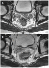
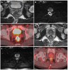
Rectal cancer is a common cancer and a major cause of mortality in Western countries. Accurate staging is essential for determining the optimal treatment strategies and planning appropriate surgical procedures to control rectal cancer. Endorectal ultrasonography (EUS) is suitable for assessing the extent of tumor invasion, particularly in early-stage or superficial rectal cancer cases. In advanced cases with distant metastases, computed tomography (CT) is the primary approach used to evaluate the disease. Magnetic resonance imaging (MRI) is often used to assess preoperative staging and the circumferential resection margin involvement, which assists in evaluating a patient's risk of recurrence and their optimal therapeutic strategy. Positron emission tomography (PET)-CT may be useful in detecting occult synchronous tumors or metastases at the time of initial presentation. Restaging after neoadjuvant chemoradiotherapy (CRT) remains a challenge with all modalities because it is difficult to reliably differentiate between the tumor mass and other radiation-induced changes in the images. EUS does not appear to have a useful role in post-therapeutic response assessments. Although CT is most commonly used to evaluate treatment responses, its utility for identifying and following-up metastatic lesions is limited. Preoperative high-resolution MRI in combination with diffusion-weighted imaging, and/or PET-CT could provide valuable prognostic information for rectal cancer patients with locally advanced disease receiving preoperative CRT. Based on these results, we conclude that a combination of multimodal imaging methods should be used to precisely assess the restaging of rectal cancer following CRT.
Keywords: Imaging; Multimodality; Rectal cancer; Restaging; Staging.
Figures


References
-
- Salerno G, Sinnatamby C, Branagan G, Daniels IR, Heald RJ, Moran BJ. Defining the rectum: surgically, radiologically and anatomically. Colorectal Dis. 2006;8 Suppl 3:5–9. - PubMed
-
- Siegel R, Naishadham D, Jemal A. Cancer statistics, 2013. CA Cancer J Clin. 2013;63:11–30. - PubMed
-
- Edge SB, American Joint Committee on Cancer. Colon and Rectum. In: AJCC cancer staging manual., editor. 7th ed. New York: Springer; 2010. pp. 143–164.
-
- Benson AB, Bekaii-Saab T, Chan E, Chen YJ, Choti MA, Cooper HS, Engstrom PF, Enzinger PC, Fakih MG, Fuchs CS, et al. Rectal cancer. J Natl Compr Canc Netw. 2012;10:1528–1564. - PubMed
Publication types
MeSH terms
LinkOut - more resources
Full Text Sources
Other Literature Sources
Research Materials

