. 2014 May;7(5):1447-1454.
doi: 10.3892/ol.2014.1950. Epub 2014 Mar 7.

Expression of astrocyte elevated gene-1 closely correlates with the angiogenesis of gastric cancer

Affiliations

Expression of astrocyte elevated gene-1 closely correlates with the angiogenesis of gastric cancer

Shuhua Li et al. Oncol Lett. 2014 May.

Abstract

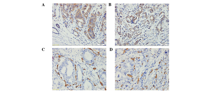
Previous studies have demonstrated that astrocyte elevated gene-1 (AEG-1) is overexpressed in several cancer types and that its upregulation may promote cell proliferation, cell transformation and tumor progression. The present study investigated the expression and prognostic value of AEG-1 in primary gastric cancer (GC) as well as its role in angiogenesis. The results obtained from real-time reverse transcription polymerase chain reaction and western blotting revealed the upregulation of AEG-1 mRNA (P=0.007) and protein expression (P<0.001) in the majority of cancerous tissues compared with matched adjacent non-cancerous gastric tissues. To further investigate the clinicopathological and prognostic roles of AEG-1, immunohistochemical analysis of 216 GC tissue blocks was performed. The results showed that high AEG-1 expression closely correlated with differentiation degree (P<0.001 ), T stage (P<0.001), N stage (P=0.003) and M stage (P=0.013). Consistent with the abovementioned results, AEG-1 upregulation was also found to significantly correlate with poor survival in GC patients (P<0.001). Furthermore, carcinomas with elevated AEG-1 expression demonstrated high vascular endothelial growth factor (VEGF) expression and microvessel density, which was labeled by cluster of differentiation 34. In addition, an AEG-1 siRNA assay in MGC-803 cells showed that the AEG-1 gene may promote VEGF and hypoxia-inducible factor-1α protein and mRNA expression. The results of the current study indicated that AEG-1 may serve as a valuable prognostic marker for GC and may be involved in regulating tumor angiogenesis.

Keywords: angiogenesis; astrocyte elevated gene-1; gastric cancer; hypoxia-inducible factor-1α; vascular endothelial growth factor.


Figures

Figure 1
Figure 1
Elevated mRNA expression of astrocyte elevated gene-1 in normal gastric mucosa and gastric cancer tissues as assessed by real-time reverse transcription polymerase chain reaction (n=20; P=0.007). Horizontal lines represent the mean and data are presented as three individually matched pairs of normal and neoplastic tissue samples.
Figure 2
Figure 2
Increased protein expression of AEG-1 in GC as assessed by western blotting. (A) Relative AEG-1 protein expression levels in GC and non-cancerous tissues (AEG-1/GAPDH; n=20; P<0.001). Horizontal lines represents the mean. (B) Representative results of AEG-1 protein expression in three pairs of gastric tumor tissues and matched adjacent non-tumorous tissues. Lane N, matched non-cancerous gastric mucosa; Lane C, GC tissues; AEG-1, astrocyte elevated gene-1; GC, gastric cancer; GAPDH, glyceraldehyde-3-phosphate dehydrogenase.
Figure 3
Figure 3
Astrocyte elevated gene-1 (AEG-1) protein expression in gastric cancer (GC) surgical specimens shown by immunohistochemistry. Immunohistochemical staining for AEG-1 was predominantly observed in the cytoplasm of the GC cells. Weak AEG-1 staining was observed in (A) the non-cancerous gastric mucosa and (B) the well-differentiated GC cells. Strong AEG-1 staining was observed in the (C) moderately and (D) poorly differentiated GC cells (stain, 3,3′-diaminobenzidine; scale bar, 100 μm; magnification, ×200).
Figure 4
Figure 4
Kaplan-Meier survival curves of gastric cancer patients (n=216). The survival rate of patients in the high astrocyte elevated gene-1 (AEG-1) expression group was significantly lower than that of patients in the low AEG-1 expression group (log-rank test; P<0.001).
Figure 5
Figure 5
Immunohistochemical staining for vascular endothelial growth factor (VEGF) is predominantly observed in the cytoplasm of lung cancer cells. (A) Strong positive expression of VEGF was observed in the moderately differentiated adenocarcinoma. (B) Positive expression of VEGF was observed in the moderately differentiated squamous carcinoma and CD34 staining was predominantly located in the surrounding microvessels of the lung carcinoma. Positive expression of CD34 was observed in the moderately differentiated (C) adenocarcinoma and (D) squamous carcinoma (streptavidin-peroxidase; scale bar, 50 μm; magnification, ×200).
Figure 6
Figure 6
Representative western blotting for AEG-1 protein expression shows downregulation of AEG-1 by siRNA. Data are presented as the means ± standard deviation of three independent experiments. *P<0.05 vs. scramble siRNA (control). AEG-1, astrocyte elevated gene-1; GAPDH, glyceraldehyde-3-phosphate dehydrogenase.
Figure 7
Figure 7
Effects of AEG-1 siRNA on VEGF and HIF-1α expression. MGC-803 cells were transfected with AEG-1 siRNA and Con-siRNA. (A) VEGF and HIF-1α were determined by western blotting. Upper panel shows a representative blot for VEGF and HIF-1α protein, and the lower panel shows the quantification of VEGF and HIF-1α protein levels. (B) VEGF and HIF-1α mRNA were determined by real-time reverse transcription polymerase chain reaction in MGC-803 cells and the levels of VEGF and HIF-1α mRNA are presented as the (VEGF and thrombospondin-1)/GAPDH mRNA ratio. Data are presented as means ± standard deviation of three independent experiments. *P<0.05 vs. Con-siRNA (control). AEG-1, astrocyte elevated gene-1; VEGF, vascular endothelial growth factor; HIF-1α, hypoxia-inducible factor-1α; Con-siRNA, scramble siRNA; GAPDH, glyceraldehyde-3-phosphate dehydrogenase.

Similar articles

Cited by

References

    1. Chen WQ. Estimation of cancer incidence and mortality in China in 2004–2005. Zhonghua Zhong Liu Za Zhi. 2009;31:664–668. (In Chinese) - PubMed
    1. Dudeja V, Habermann EB, Zhong W, et al. Guideline recommended gastric cancer care in the elderly: insights into the applicability of cancer trials to real world. Ann Surg Oncol. 2011;18:26–33. - PubMed
    1. Park MO, Park HA. Development of a nursing practice guideline for pre and post-operative care of gastric cancer patients. Healthc Inform Res. 2010;16:215–223. - PMC - PubMed
    1. Chen CN, Lin JJ, Chen JJ, et al. Gene expression profile predicts patient survival of gastric cancer after surgical resection. J Clin Oncol. 2005;23:7286–7295. - PubMed
    1. Kang DC, Su ZZ, Sarkar D, et al. Cloning and characterization of HIV-1-inducible astrocyte elevated gene-1, AEG-1. Gene. 2005;353:8–15. - PubMed

LinkOut - more resources