A case of idiopathic encephalomeningocele
- PMID: 24765291
- PMCID: PMC3981251
- DOI: 10.4081/cp.2011.e29
A case of idiopathic encephalomeningocele
Abstract
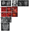
In the present case we report about an encephalomeningocele in an adult female. Since the cause of this medical entity is a congenital fusion defect of the neural tube of the cranial base, most of the encephaloceles occurs in children leading to facial disfigurement. In the rare cases described in adults, rhinorrhea is usually present. Here we present a case of temporobasal encephalomeningocele in a 72-year-old female patient suffering from headaches in the last 4-5 years. No rhinorrhea or other significant neurological symptoms were noticed. No congenital cause was apparent. After diagnostic steps including brain magnetic resonance imaging (MRI), cranial computed tomography (CT) and MR cisternography, an encephalomeningocele was diagnosed. Through a pterional approach this was completely removed. The only symptom the patient complaint about, headache, was eliminated after surgery.
Keywords: congenital defect; headache.; neurosurgical removal; temporobasal encephalomeningocele.
Figures

References
-
- Bozinov O, Tirakotai W, Sure U, Bertalanffy H. Surgical closure and reconstruction of a large occipital encephalocele without parenchymal excision. Childs Nerv Syst. 2005;21:144–7. - PubMed
-
- Vannouhuys JM, Bruyn GW. Nasopharyngeal transsphenoidal encephalocele, craterlike hole in the optic disk, and agenesis of the corpus callosum. Pneumoencephalographic visualization in a case. Psychiat Neurol Neurochir. 1964;67:243–58. - PubMed
-
- Garg P, Rathi V, Bhargava SK, Aggarwal A. CSF Rhinorrhea and recurrent meningitis caused by transethmoidal meningoencephaloceles. Indian Pediatr. 2005;42:1033–6. - PubMed
-
- Kishikawa K, Nagao T, Ibayashi S, et al. [An adult case of basal encephalomeningocele with recurrent meningitis] Rinsho Shinkeigaku. 1994;34:908–10. - PubMed
-
- Nishikawa T, Ishida H, Nibu K. A rare spontaneous temporal meningoencephalocele with dehiscence into the pterygoid fossa. Auris Nasus Larynx. 2004;31:429–31. - PubMed
Publication types
LinkOut - more resources
Full Text Sources

