

Post-traumatic extensive knee ganglion cyst
- PMID: 24765322
- PMCID: PMC3981388
- DOI: 10.4081/cp.2011.e61
Post-traumatic extensive knee ganglion cyst
Abstract
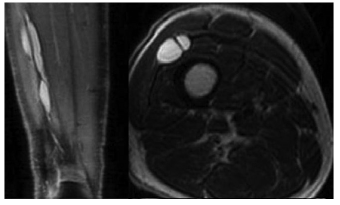
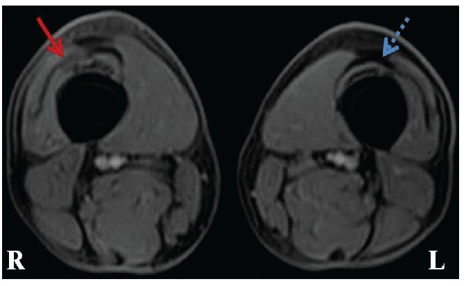
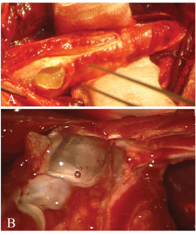
A rare case of a posttraumatic extensive ganglion cyst of the anterolateral thigh with connection to the knee joint is presented. A 54-year-old man presented a palpable mass in the anterolateral region of his right thigh with a 15 months existing sense of fullness and tightness. He had an accident with his bicycle 21 months ago. Magnetic resonance imaging (MRI) was performed showing a cyst inside the quadriceps femoris muscle between vastus lateralis and intermedius with connection to recessus suprapatellaris and knee joint. In addition MRI detected a traumatic lesion in the quadriceps femoris tendon in the near of the knee joint. The ganglion cyst was 18 cm long and was excised completely. Intraoperatively, the knee joint connection was confirmed and excised as well. The ganglion cyst was filled with a gelatinous and viscous fluid.
Keywords: ganglion cyst; knee joint.; post-traumatic; quadriceps femoris tendon.
Figures
Similar articles
-
Giant ganglion cyst of the quadriceps femoris tendon.Knee Surg Sports Traumatol Arthrosc. 2003 Jul;11(4):260-2. doi: 10.1007/s00167-003-0364-9. Epub 2003 May 10. Knee Surg Sports Traumatol Arthrosc. 2003. PMID: 12740653
-
New insight in the architecture of the quadriceps tendon.J Exp Orthop. 2016 Dec;3(1):32. doi: 10.1186/s40634-016-0068-y. Epub 2016 Nov 3. J Exp Orthop. 2016. PMID: 27813020 Free PMC article.
-
MR imaging of the quadriceps femoris tendon: distal tear characterization and clinical significance of rupture types.Eur Radiol. 2021 Oct;31(10):7674-7683. doi: 10.1007/s00330-021-07912-y. Epub 2021 Apr 16. Eur Radiol. 2021. PMID: 33860830 Free PMC article.
-
Intratendinous ganglion cyst of the semimembranosus tendon.Br J Radiol. 2010 Apr;83(988):e79-82. doi: 10.1259/bjr/23178227. Br J Radiol. 2010. PMID: 20335437 Free PMC article. Review.
-
An intraneural ganglion cyst of the ulnar nerve at the wrist: a case report and literature review.J Int Med Res. 2021 Jan;49(1):300060520982701. doi: 10.1177/0300060520982701. J Int Med Res. 2021. PMID: 33459091 Free PMC article. Review.
Cited by
-
Tibial Component Revision Arthroplasty Using Porous Tantalum Cone for Symptomatic Progressive Periprosthetic Proximal Tibial Ganglion Cyst about All-Polyethylene Tibia Primary Total Knee Replacement: A Case Report and Review of Literature.J Orthop Case Rep. 2024 Feb;14(2):131-135. doi: 10.13107/jocr.2024.v14.i02.4244. J Orthop Case Rep. 2024. PMID: 38420232 Free PMC article.
-
Ganglion cyst at the proximal tibiofibular joint - A rare cause of compression neuropathy of the peroneal nerve.Radiol Case Rep. 2021 Nov 2;17(1):99-102. doi: 10.1016/j.radcr.2021.10.004. eCollection 2022 Jan. Radiol Case Rep. 2021. PMID: 34765070 Free PMC article.
References
-
- Burk DL, Jr, Dalinka MK, Kanal E, et al. Meniscal and ganglion cysts of the knee: MR evaluation. AJR Am J Roentgenol. 1988;150:331–6. - PubMed
-
- Fukuda A, Kato K, Sudo A, Uchida A. Ganglion cyst arising from the posterolateral capsule of the knee. J Orthop Sci. 2010;15:261–4. - PubMed
-
- Vayvada H, Tayfur V, Menderes A, et al. Giant ganglion cyst of the quadriceps femoris tendon. Knee Surg Sports Traumatol Arthrosc. 2003;11:260–2. - PubMed
-
- Lakdawala A, Ireland J. Giant ganglion formation in the quadriceps femoris tendon following total knee replacement. Int J Clin Pract. 2004;58:990–1. - PubMed
-
- Vilchez F, Erquicia J, Pelfort X, Monllau JC. [Symptomatic ganglions in the knee joint. Report of three cases and literature review] Acta Ortop Mex. 2009;23:223–7. - PubMed
Publication types
LinkOut - more resources
Full Text Sources
