Case Reports
. 2011 Sep 30;1(3):e66.
doi: 10.4081/cp.2011.e66. eCollection 2011 Jul 1.

Hydrocephalus, a rare manifestation of sarcoidosis

Affiliations
Case Reports

Hydrocephalus, a rare manifestation of sarcoidosis

Johan M van Rooijen et al. Clin Pract. .

Abstract

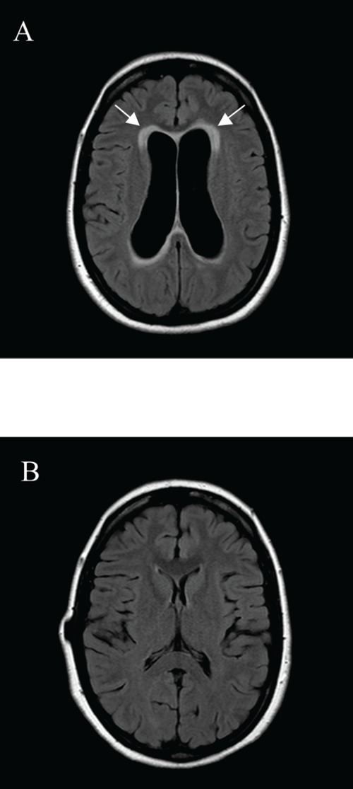
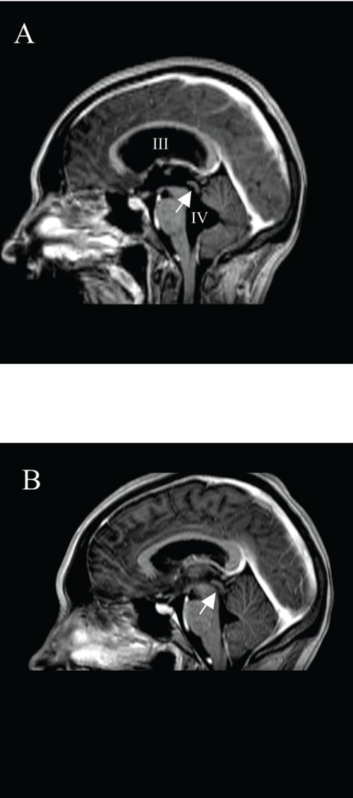
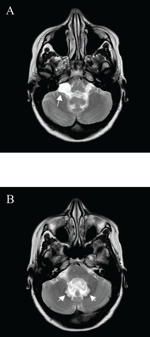
A 36-week-pregnant woman developed a symptomatic hydrocephalus. Chest imaging showed bihilar lymphadenopathy and histological examination of a mediastinal lymph node revealed non-caseating granulomas. After delivery, her neurologic complaints progressed. Placement of a ventriculoperitoneal drain (VPD) did not reduce the symptoms. However, steroids resulted in rapid disappearance of the hydrocephalus. Hydrocephalus is a very rare manifestation of sarcoidosis. The diagnosis relies on the ability of clinicians to recognize this disorder. This case shows how a difference in opinion of the several specialists involved can lead to a delay in diagnosis and treatment.

Keywords: hydrocephalus; neurosarcoidosis.; sarcoidosis.


Figures

Figure 1
Figure 1
Magnetic resonance imaging of the head, axial FLAIR images at the level of the lateral ventricles. (A) First scan at presentation, showing a dilated ventricular system accompanied with periventricular transependymal effusion of CSF (arrows). (B) Placement of a VPD did not change the hydrocephalus, but after three weeks of therapy with steroids normalization of the ventricles width and resolution of the transependymal effusion of CSF is demonstrated.
Figure 2
Figure 2
Magnetic resonance imaging of the head, median sagittal T1 weighted images post contrast injection. (A) Dilated third en fourth ventricles and dilated aquaduct (arrow) can be appreciated. There are no signs of pathological ependymal enhancement or enhancing masses.(B) Placement of a VPD did not change the hydrocephalus, but after three weeks of therapy with steroids, normal size and aspect of the third and fourth ventricle and aquaduct (arrow) is observed.
Figure 3
Figure 3
Magnetic resonance imaging of the head, axial T2 weighted images at the level of posterior fossa. First scan at presentation, showing (A) enlargement of the cerebellomedullary cisterns. The signal intensities of the CSF (arrow) suggest a membranous outflow-obstruction at the foramina of Luschka, although no cerebral mass lesions and no obstruction could be demonstrated. (B) This image shows an enlarged fourth ventricle and a significant transependymal effusion of CSF dorsally (arrows), a phenomenon not seen very often in the posterior fossa.

Similar articles

Cited by

References

    1. Spencer TS, Campellone JV, Maldonado I, et al. Clinical and magnetic resonance imaging manifestations of neurosarcoidosis. Semin Arthritis Rheum. 2004;34:649–61. - PubMed
    1. Lower EE, Broderick JP, Brott TG, et al. Diagnosis and management of neurological sarcoidosis. Arch Intern Med. 1997;157:1864–8. - PubMed
    1. Akhondi H, Barochia S, Holmström B, Williams MJ. Hydrocephalus as a presenting manifestation of neurosarcoidosis. Southern Med J. 2003;96:403–6. - PubMed
    1. Benzagmout M, Boujraf S, Góngora-Rivera F, et al. Neurosarcoidosis which manifested as acute hydrocephalus: diagnosis and treatment. Intern Med. 2007;46:1601–4. - PubMed
    1. Lee CH, Jung YS, Lee SH. Hydrocephalus as a presenting manifestation of neurosarcoidosis: easy to misdiagnose as tuberculosis. J Korean Neurosurg Soc. 2010;48:79–81. - PMC - PubMed

Publication types

LinkOut - more resources