

Coronary artery anomalies presenting with ST-segment elevation myocardial infarction
- PMID: 24765348
- PMCID: PMC3981407
- DOI: 10.4081/cp.2011.e107
Coronary artery anomalies presenting with ST-segment elevation myocardial infarction
Abstract
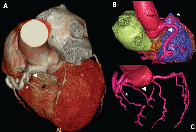
ST-segment elevation MI (STEMI) is a rare presentation in patients with coronary artery anomalies. In these patients, the identification of the culprit lesion and its treatment may be difficult, particularly in the emergency setting of primary percutaneous coronary intervention (PCI). From January 2008 to April 2011, 1015 STEMI patients received coronary artery angiography and primary PCI in our centre. Of these, 5 (0.4%) patients showed a coronary artery anomaly. In this paper we reported two rare cases: i) the first is a single coronary artery originating from right sinus of Valsalva; ii) the second is a separate origin of 3 coronary arteries originating from the right sinus of Valsalva. In conclusion, coronary artery anomalies presenting with STEMI are really uncommon, but often are a challenge. The integration between traditional coronary artery angiography and multidetector computerized tomography is crucial to optimize the interventional and medical management of these patients.
Keywords: coronary artery anomalies; multidetector computerized tomography.; myocardial infarction; primary percutaneous coronary intervention.
Figures


References
-
- Alexander RW, Griggith GC. Anomalies of the coronary arteries and their clinical significance. Circulation. 1956;14:800–5. - PubMed
-
- Yamanaka O, Hobbs RE. Coronary artery anomalies in 126,595 patients undergoing coronary arteriography. Cathet Cardiovasc Diagn. 1990;21:28–40. - PubMed
-
- Angelini P, Velasco JA, Flamm S. Coronary anomalies: incidence, pathophysiology, and clinical relevance. Circulation. 2002;105:2449–54. - PubMed
-
- Angelini P. Coronary artery anomalies: an entity in search of an identity. Circulation. 2007;115:1296–3. - PubMed
-
- Maron BJ, Carney KP, Lever HM, et al. Relationship of race to sudden cardiac death in competitive athletes with hypertrophic cardiomyopathy. J Am Coll Cardiol. 2003;41:974–80. - PubMed
Publication types
LinkOut - more resources
Full Text Sources
Miscellaneous
