Disseminated herpes zoster ophthalmicus in an immunocompetent 8-year old boy
- PMID: 24765504
- PMCID: PMC3981262
- DOI: 10.4081/cp.2013.e16
Disseminated herpes zoster ophthalmicus in an immunocompetent 8-year old boy
Abstract
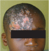
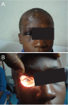
Varicella results from a primary infection with the varicella virus while herpes zoster is caused by a reactivation of a latent infection. Dissemination of herpes zoster is uncommon in immunocompetent individuals. Reports of disseminated herpes zoster in children are even less common than in adults. An unusual case of disseminated herpes zoster ophthalmicus in an 8-year old immunocompetent black boy is presented. He had a previous primary Varicella zoster virus infection at three years of age. In the current report, he presented during an on-going chicken pox outbreak and survived with no significant complications. A breakthrough varicella virus re-infection or a reactivation is possible, both of which could present as zoster. This case emphasizes the need for prevention of varicella virus infection through universal childhood immunization and effective infection control strategies in health care settings.
Keywords: child; disseminated; herpes zoster; immunocompetent.
Figures


Similar articles
-
The Skin and the Eye - Herpes Zoster Ophthalmicus in a Healthy 18-month-old Toddler.Folia Med (Plovdiv). 2018 Mar 1;60(1):170-174. doi: 10.1515/folmed-2017-0064. Folia Med (Plovdiv). 2018. PMID: 29668453
-
Pediatric Herpes Zoster in a 10-Year-Old Boy With Delayed Immunizations: A Case Report.Cureus. 2025 Mar 18;17(3):e80787. doi: 10.7759/cureus.80787. eCollection 2025 Mar. Cureus. 2025. PMID: 40255789 Free PMC article.
-
Herpes Zoster Ophthalmicus, Central Retinal Artery Occlusion, and Neovascular Glaucoma in an Immunocompetent Individual.J Ophthalmic Vis Res. 2019 Jan-Mar;14(1):97-100. doi: 10.4103/jovr.jovr_65_17. J Ophthalmic Vis Res. 2019. PMID: 30820294 Free PMC article.
-
[Reactivation of herpes zoster infection by varicella-zoster virus].Med Pregl. 1999 Mar-May;52(3-5):125-8. Med Pregl. 1999. PMID: 10518396 Review. Croatian.
-
Disseminated varicella-zoster virus in an immunocompetent adult.Dermatol Online J. 2015 Feb 22;21(3):13030/qt3cz2x99b. Dermatol Online J. 2015. PMID: 25780980 Review.
Cited by
-
Pediatric herpes zoster ophthalmicus: a systematic review.Graefes Arch Clin Exp Ophthalmol. 2023 Aug;261(8):2169-2179. doi: 10.1007/s00417-023-06033-0. Epub 2023 Mar 23. Graefes Arch Clin Exp Ophthalmol. 2023. PMID: 36949170 Free PMC article.
-
Disseminated Zoster Involving the Whole Body in an Immunocompetent Patient Complaining of Left Leg Radiating Pain and Weakness: A Case Report and Literature Review.Geriatr Orthop Surg Rehabil. 2022 Aug 10;13:21514593221119619. doi: 10.1177/21514593221119619. eCollection 2022. Geriatr Orthop Surg Rehabil. 2022. PMID: 35983318 Free PMC article.
-
Disseminated Herpes Zoster on a Child with Systemic Lupus Erythematosus and Lupus Nephritis.Infect Drug Resist. 2021 Jul 20;14:2777-2785. doi: 10.2147/IDR.S314220. eCollection 2021. Infect Drug Resist. 2021. PMID: 34321894 Free PMC article.
-
Bilateral symmetrical herpes zoster in an immunocompetent 15-year-old adolescent boy.Case Rep Pediatr. 2015;2015:121549. doi: 10.1155/2015/121549. Epub 2015 Jan 27. Case Rep Pediatr. 2015. PMID: 25692062 Free PMC article.
-
Herpes zoster with cutaneous dissemination: a rare presentation of an uncommon pathology in children.BMJ Case Rep. 2018 Jun 12;2018:bcr2018225355. doi: 10.1136/bcr-2018-225355. BMJ Case Rep. 2018. PMID: 29895579 Free PMC article.
References
-
- Myers MG, Seward JF, LaRussa PS.Varicella-zoster virus. Kliegman RM, Behrman RE, Jenson HB, Stanton BF, Nelson textbook of paediatrics. 19th edition Philadelphia, PA: Saunders; 2011. pp 1104-1105
-
- Sterling JC.Virus infections. Burns T, Breathnach S, Cox N, Griffths C, Rooks textbook of dermatology. Vol. 2 8th edition London: Wiley-Blackwell; 2011. p 33.25
-
- Brown TJ, McCrary M, Tyring SK.Varicella-zoster virus (Herpes 3). J Am Acad Dermatol. 2002; 46:972-97 - PubMed
-
- Wolff K, RA J.Viral infections of skin and mucosa. In: EDITORS??Fitzpatrick’s color atlas and synopsis of clinical dermatology. 6th edition NewYork: McGraw Hill; 2009. pp 837-45
Publication types
LinkOut - more resources
Full Text Sources
Other Literature Sources
