. 2011 Dec 28;1(1):20-32.

Inhibition of Rab5 Activation During Insulin Receptor-Mediated Endocytosis

Affiliations

Inhibition of Rab5 Activation During Insulin Receptor-Mediated Endocytosis

Ivan Jozic et al. Curr Cell Biochem. .

Abstract

Activation of receptor tyrosine kinases is a key feature in receptor signaling and membrane trafficking processes. In this study, we found that the insulin receptor tyrosine kinase activity is required for fusion between early endosomes. AG1024, a receptor tyrosine kinase inhibitor, blocked the in vitro endosome fusion in a concentration-dependent manner. We observed that Rab5: wild type partially rescued the fusion reaction, whereas Rab5: Q79L mutant fully rescued it. We also observed that treatment of cells with insulin receptor kinase inhibitor HNMPA-(AM)3 blocked the formation of Rab5-positive endosomes as well as the activation of Rab5 upon addition of insulin in intact cells. HNMPA-(AM)3 inhibitor also affected the endosomal co-localization of Rab5 and insulin receptor. However, the formation of Rab5: Q79L mutant-positive endosomes were not affected by the HNMPA-(AM)3 inhibitor. In addition, HNMPA-(AM)3 inhibitor affected the association of Rin1 to membrane upon insulin stimulation. Furthermore, Rin1 did not fully support endosome fusion in the presence of the AG1024 inhibitor. These results constitute the first evidence that, at least in part, the enzymatic activity of insulin receptor is required for the fusion events via the activation of Rab5.

Keywords: Endosome fusion; Kinase Inhibitors; Receptor tyrosine kinase; small GTPases.


Figures

Figure 1
Figure 1
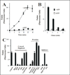
Effect of insulin receptor kinase inhibitor on fusion between endosomes. (A) Effect of cytosol and energy on the endosome-endosome fusion. Five-min vesicles containing either Biotin-Insulin (B-Ins) or Avidin-β-Galactosidase (Av-GAL) were mixed in fusion buffer supplemented with different amount of cytosol containing ATP-regenerated (■) or ATP-depleted systems (●). Samples were then transferred to 37°C for the indicated times and processed as described in Material and Methods to determine the percentage forming the immune complex formation. The data are presented as means ± SD of four independent experiments. (B) Fusion assay was performed under standard conditions as described above either in the absence (□) or in the presence of different amounts of AG9 (○) and AG1024 (●) containing 1 mg/ml of cytosol. The data are presented as means ± SD of four independent experiments.
Figure 2
Figure 2
Fusion between endosomes. (A) Effect of time on the endosome-endosome fusion. Two sets of vesicles prepared from cells that have been allowed to endocytose B-Ins and Av-GAL for 5 min at 37°C, respectively. Then the vesicles were mixed in fusion buffer supplemented with different amounts of cytosolic proteins /ml containing ATP-regenerated (●) or ATP-depleted systems (■). Samples were then transferred to 37°C for the indicated times and processed as described in Material and Methods to determine the percentage immune complex formation. The data are presented as means ± SD of four independent experiments. (B) Av-GAL was internalized by a 3-min uptake at 37°C and chased for 0, 3, 6, 12 min. Cells were then incubated at 4°C for 1 h with B-Ins. Endocytosis of Insulin was induced by incubation at 37°C and the amount of B-Ins associated with Av-GAL was quantitated at each time by solubilizing the cells in the presence of excess of biotin-BSA. Results are expressed as fluorescence units, which indicates the percentage of the immune complex formed after solubilization of vesicles in the absence of biotin-BSA. (C) Vesicles fractions were obtained and resuspended in homogenization and treated as follow: Antibodies: 200 ng/ml control IgG, or anti-Rab4, anti-Rab5, or anti-EEA1, or anti-Rab11, or anti-Rin1 antibodies were added to fusion reaction; GDI proteins: vesicles were incubated in the presence of 1 uM GDI, washed and then resuspended in fusion buffer; Rab5 proteins: 0.3 µg/ml Rab5-REP-1 complex was added to the fusion reaction; Rin1 proteins: 0.2 µg/ml of Rin1 was added to the fusion reaction; NEM treatment: Cytosol and vesicles were pretreated with 0.75 mM NEM at 4°C for 45 min and excess of NEM was inactivated by 2 mM DTT. Fusion was carried out in the presence of ATP-regenerating system, and the ATP-dependent fusion was expressed as a fraction of the fusion obtained with non-treated vesicles resuspended in 1.5 mg of normal cytsosol/ml for 45 min at 37°C. Background was measured by incubation for 45 min at 4°C. The data are presented as means ± SD of three independent experiments.
Figure 3
Figure 3. Morphology of in vitro fusion between endosomes
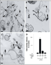
Sub-cellular fractions containing colloidal gold of 10 nm (B-Ins) and 20 nm (Av-GAL) loaded in 5-min early endosomes were incubated in vitro for 45 min in fusion buffer in the presence of 1.2 mg of cytosolic protein/ml at 37°C (A) and at 4°C (B). (C) Fuison was also supplemented by 20 µM AG1024 kinase inhibitor in the presence of 1.2 mg of cytosolic protein/ml at 37°C. After the fusion, the samples were fixed in suspension, pelleted, and analyzed for transmission electron microscopy. Bars: 100 nm. (D) For each time of the fusion reaction, the presence of either 10 or 20 nm gold particles was assessed for at least 2,000 endosomes under four different experimental conditions (−ATP/37°C, lane 1; +ATP/37°C, lane 2; +ATP/37°C in the presence of 20 µM AG1024 (KI), lane 3; +ATP/4°C, lane 4). Results are expressed as a percentage of the total endosomes that contained 10 nm and 20 nm gold. Values are means ± SD, n = 3.
Figure 4
Figure 4
Selective effect of insulin receptor kinase inhibitor on the fusion reaction. The fusion assay was performed under standard conditions as described in Figure 1A, either in the absence or in the presence of Rab5:WT or Rab5:Q79L mutant, supplemented with either 20 µM AG1024 or ATP in the presence of 0.5 mg/ml cytosol. The data are presented as means ± SD of four independent experiments.
Figure 5
Figure 5
Insulin receptor kinase inhibitor blocks endosome fusion stimulated by Rin1. The fusion assay was performed under standard conditions as described in Figure 1A, either in the absence or in the presence of different concentrations of Rin1 supplemented with several concentrations of AG1024 (○: no addition, ●: 20 µM) inhibitor in the presence of 0.5 mg/ml cytosol. The data are presented as means ± SD of four independent experiments.
Figure 6
Figure 6
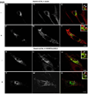
Confocal immunofluorescence analysis of cells co-expressing Insulin receptor, Rab5: wild-type in the presence of insulin receptor tyrosine kinase inhibitor. NIH-IR cells were transfected with plasmids encoding GFP-Rab5: wild type (A–L) in the absence (A–C; G–I) or in the presence of 100 ng/ml insulin (D–F; J–L). Cells were also supplemented with 100 µM HNMPA-(AM)3 inhibitors (G–L) and inactive analog (AG9) (A–F). 100 ng/ml insulin was bound to the cells at 4°C for 60 min. The cells were then washed with ice-cold PBS and then incubated at 37°C for 8 min. Subsequently, the cells were washed three times with ice-cold PBS and fixed with 4% paraformaldehyde and then were permeabilized with 0.1% Triton X-I00 prior to incubation with antibodies. The primary antibodies used were rabbit anti-insulin receptor. The secondary antibodies used were Alexa564-labelled donkey anti-rabbit IgG antibodies. Yellow color indicates areas of co-localization between the internalized insulin receptor and the overexpressed GFP-Rab5 proteins (C, F, I and L). An inactive analog (AG9) was used as control. The optical sections viewed are 0.4 µm. Size bars, 10 µm.
Figure 7
Figure 7
Confocal immunofluorescence analysis of cells co-expressing Insulin receptor, Rab5: Q79L mutant in the presence of insulin receptor tyrosine kinase inhibitor. NIH-IR cells were transfected with plasmids encoding GFP-Rab5: Q79L (A–L) and in the absence (A–C and G–I) or in the presence of insulin (D–F and J–L). Cells were also supplemented with 100 µM HNMPA-(AM)3 inhibitors (G–L) and inactive analog (AG9) (A–F). 100 ng/ml insulin was bound to cells at 4°C for 60 min. Cells were washed with ice-cold PBS and then incubated at 37°C for 8 min. Subsequently, the cells were washed three times with ice-cold PBS and fixed with 4% paraformaldehyde and then permeabilized with 0.1% Triton X-I00 prior to incubation with antibodies. The primary antibodies used were rabbit anti-insulin receptor. The secondary antibodies used were Alexa564-labelled donkey anti-rabbit IgG antibodies. Yellow color indicates areas of co-localization between the internalized insulin receptor and the overexpressed GFP-Rab5 proteins (C, F, I and L). An inactive analog (AG9) was used as control. The optical sections viewed are 0.4 µm. Size bars, 10 µm.
Figure 8
Figure 8
Insulin receptor tyrosine kinase inhibitor blocks the activation of Rab5 in intact cells. NIH-IR were transfected with plasmids encoding GFP-Rab5: wild type (A) and GFP-Rab5: Q79L mutant (B) in the absence or in the presence of 100 ng/ml insulin containing either 100 µM AG9 or 100 µM HNMPA-(AM)3 (HNM) inhibitors as indicated in the Figure. Insulin was bound to the cells at 4°C for 60 min. The cells were washed and then incubated at 37°C for 8 min. Subsequently, the cells were washed three times with ice-cold PBS, lysed and incubated with gluthathione beads either in the presence of GST alone or GST-EEA1 at 4°C for 60 min. After incubation, the beads were washed and the presence of activated Rab5 was analyzed by Western blotting using anti-Rab5 antibodies. The data are presented as means ± SD of four independent experiments.
Figure 9
Figure 9
Effect of HNMPA-(AM)3 inhibitor on the tyrosine phosphorylation of Insulin receptor and recruitment of Rin1. Cells were incubated in the absence or in the presence of 100 ng/ml insulin either 100 µM AG9 or 100 µM HNMPA-(AM)3 inhibitor as described in the Figure. After treatment, cells were washed with ice cold PBS and incubated at 37°C for 8 minutes. Cells were then washed again using ice-cold PBS, homogenized and membrane fractions were prepared as described in Materials and Methods. Membrane (M) and cytosol (S) fractions were treated with sample buffer and proteins were subject to SDS-PAGE, blotted to a nitrocellulose membrane, and antibodies specific to IRS-1 (A), Grb2 (B), Rin1 (B), Rabex-5 (C), tubulin, phospo-insulin receptor (p)-IR and total (t)-IR, and tubulin were used to visualize these proteins by Western blot analysis. A representative experiment is shown (E). This experiment was repeated at least three times, and the results were reproducible. The data are presented as means ± SD of three independent experiments.
Figure 10
Figure 10
Activation of Rab5 during fluid phase and receptor-mediated endocytosis. NIH-IR cells were transfected with plasmids encoding GFP-Rab5: wild type in the absence or in the presence of 100 ng/ml insulin (INS), 2 mg/ml HRP and 1 ug/ml β-Galactosidase (GAL) as indicated in the Figure. Each ligand was incubated with cells at 4°C for 60 min. Cells were then incubated at 37°C for 8 min. Subsequently, the cells were washed three times with ice-cold PBS, lysed and incubated with glutathione beads either in the presence of GST-EEA1 at 4°C for 60 min. After incubation, the beads were washed and the presence of activated Rab5 was analyzed by Western blotting using anti-Rab5 antibodies. The data are presented as means ± SD of two independent experiments.

Similar articles

Cited by

References

    1. Backer JM, Shoelson SE, Haring E, White MF. Insulin receptors internalize by a rapid, saturable pathway requiring receptor autophosphorylation and an intact juxtamembrane region. Journal of Cell Biology. 1991;115:1535–1545. - PMC - PubMed
    1. Barbieri MA, Hoffenberg S, Roberts R, Mukhopadhyay A, Pomrehn A, Dickey BF, Stahl PD. Evidence for a symmetrical requirement for Rab5-GTP in in vitro endosome-endosome fusion. Journal of Biological Chemistry. 1998;273:25850–25855. - PubMed
    1. Barbieri MA, Li G, Colombo MI, Stahl PD. Rab5, an early acting endosomal GTPase, supports in vitro endosome fusion without GTP hydrolysis. Journal of Biological Chemistry. 1994;269:18720–18722. - PubMed
    1. Barbieri MA, Roberts RL, Gumusboga A, Highfield H, Alvarez-Dominguez C, Wells A, Stahl PD. Epidermal growth factor and membrane trafficking. EGF receptor activation of endocytosis requires Rab5a. Journal of Cell Biology. 2000;151:539–550. - PMC - PubMed
    1. Braell WA. Fusion between endocytic vesicles in a cell-free system. Proceedings of the National Academy of Sciences U S A. 1987;84:1137–1141. - PMC - PubMed

LinkOut - more resources