. 2014 Sep-Dec;28(3-4):374-88.
doi: 10.3109/01677063.2014.891590. Epub 2014 Apr 28.

Multiple redundant medulla projection neurons mediate color vision in Drosophila

Affiliations

Multiple redundant medulla projection neurons mediate color vision in Drosophila

Krishna V Melnattur et al. J Neurogenet. 2014 Sep-Dec.

Abstract

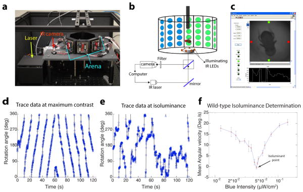
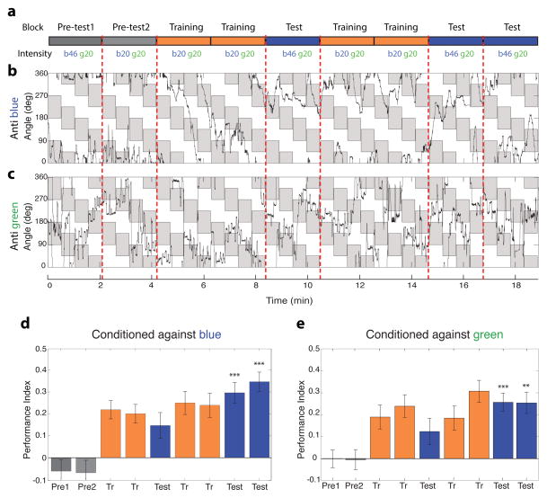
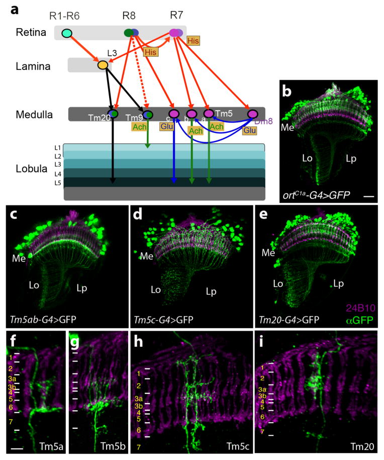
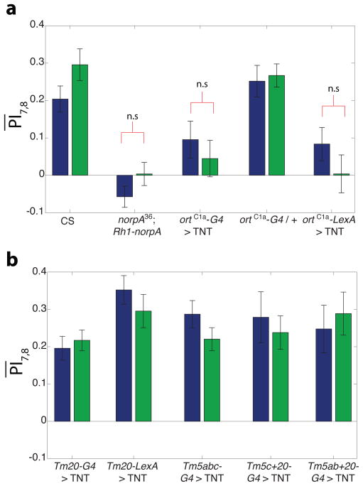
The receptor mechanism for color vision has been extensively studied. In contrast, the circuit(s) that transform(s) photoreceptor signals into color percepts to guide behavior remain(s) poorly characterized. Using intersectional genetics to inactivate identified subsets of neurons, we have uncovered the first-order interneurons that are functionally required for hue discrimination in Drosophila. We developed a novel aversive operant conditioning assay for intensity-independent color discrimination (true color vision) in Drosophila. Single flying flies are magnetically tethered in an arena surrounded by blue and green LEDs (light-emitting diodes). The flies' optomotor response is used to determine the blue-green isoluminant intensity. Flies are then conditioned to discriminate between equiluminant blue or green stimuli. Wild-type flies are successfully trained in this paradigm when conditioned to avoid either blue or green. Functional color entrainment requires the function of the narrow-spectrum photoreceptors R8 and/or R7, and is within a limited range, intensity independent, suggesting that it is mediated by a color vision system. The medulla projection neurons, Tm5a/b/c and Tm20, receive direct inputs from R7 or R8 photoreceptors and indirect input from the broad-spectrum photoreceptors R1-R6 via the lamina neuron L3. Genetically inactivating these four classes of medulla projection neurons abolished color learning. However, inactivation of subsets of these neurons is insufficient to block color learning, suggesting that true color vision is mediated by multiple redundant pathways. We hypothesize that flies represent color along multiple axes at the first synapse in the fly visual system. The apparent redundancy in learned color discrimination sharply contrasts with innate ultraviolet (UV) spectral preference, which is dominated by a single pathway from the amacrine neuron Dm8 to the Tm5c projection neurons.

Keywords: color discrimination; medulla projection neurons; neural substrate; visual behavior.


Figures

Figure 1
Figure 1. An aversive operant conditioning paradigm for true color vision
(a) Photograph of the flight simulator. An octagonal arena with blue (470nm) and green (528nm) LEDs allows for display of versatile visual stimuli, an infra-red camera is used to monitor the fly’s position, and a near-infra-red laser is used to condition the fly with a heat beam. (b) Schematic of a tethered fly in the arena. A mirror and beam splitter below the stage allow for a heat beam from the IR laser to be directed to the fly’s abdomen and for reflected illuminating light from the fly to be directed to the camera. Custom software written in LabVIEW (screenshot in c) controls display of visual patterns, monitors the fly’s position and conditions the fly when appropriate. (d–f) Using the fly’s optomotor response to determine the point of blue-green isoluminance. The fly’s angular orientation is plotted as a function of time to generate a trace of the fly’s optomotor response at maximum contrast (d) and at the isoluminant point (e). Flies are shown rotating patterns of alternating green bars of fixed intensity and blue bars of variable intensity. Plotting the average angular velocity of flies as a function of blue intensity (f), allows for determination of the blue-green isoluminant point, as the intensity that elicits minimal average angular velocity. Error bars = ±1SEM.
Figure 2
Figure 2. Blue-green color entrainment requires the functions of the inner photoreceptors R8 and/or R7
(a) Trace data of wild-type flies conditioned against blue. Flies are shown a stimulus consisting of alternating equiluminant blue and green quadrants which are rotated by 45° every 30s. The fly’s angular orientation is plotted as function of time, quadrants where the fly is punished are shaded in grey. In this experiment the grey blocks in the figure correspond to green quadrants in the arena, and white blocks to blue quadrants. The rotation of the quadrants is thus represented by the periodic change in orientation of the grey patches. The conditioning regimen is schematized above, the laser is turned on in the training blocks to condition flies, but kept off in the test and pre-test blocks. (b) Trace data of Rh1-rescued flies conditioned against blue. R1-R6 function was rescued by expressing the phospholipase C norpA under the control of a Rh1 promoter, in a norpA null mutant background. (c–f) Bar plots of the performance index (PI) of wild-type (c,d) and Rh1-rescued flies (e,f) conditioned against blue (c,e) or green (d,f). In contrast to wild-type flies, performance of Rh1-rescued flies in the test periods was not significantly different from pre-test indicating that Rh1 function is not sufficient for color entrainment. Bars are color coded as per the schematic in (a). PI of the test blocks was compared to the pre-test by a two-tailed t-test. *** p<0.001, ** p<0.01, n.s. not significant (p>0.05). Average of the two test blocks was computed ( PI7,8¯) for each condition. Genotypes: CantonS (wild-type), norpA36; Rh1-norpA (Rh1 rescue). n=19–21 flies per condition. Error bars = ±1SEM.
Figure 3
Figure 3
Color entrainment is intensity independent in a two-fold intensity range. (a) Schematic of the training paradigm for testing intensity independence. Flies were trained at equiluminant blue and green intensities (‘b20 g20’), but tested at a brighter blue intensity (‘b46 g20’). (b–c) Trace data of wild type (CantonS) flies conditioned against blue (b) or green (c) in the intensity independence paradigm. Visual stimulus display is as in Figure 2a. As in Figure 2a, the fly’s angular orientation is plotted as function of time, quadrants where the fly is punished are shaded in grey. Grey shaded blocks in (b) correspond to green quadrants in the arena, and to blue quadrants in (c). (d–e) Bar plots of wild-type flies conditioned against blue (d) or green (e). Flies retained the ability to discriminate blue from green when they were challenged with the brighter blue, indicating that learning is intensity independent in this intensity range. PI of the test blocks was compared to the pre-test for the higher blue intensity by a two-tailed t-test. *** p<0.001, ** p<0.01
Figure 4
Figure 4. Visual circuits downstream of chromatic photoreceptors
(a) Schematic of currently known visual circuits, highlighting the neurons known to receive input from multiple photoreceptors. Neuronal cell bodies are denoted by ovals. Arrows indicate axonal projections, a solid arrow between two neurons indicates a chemical synapse validated by electron microscopy, a dashed arrow indicates a chemical synapse determined using GRASP (GFP Reconstitution Across Synaptic Partners; Feinberg et al.,2007; Gordon and Scott, 2009; Karuppudurai et al., 2014). Arrows are color coded according to the neurotransmitter system expressed in the particular neuronal type. Thus, histaminergic projections from the photoreceptors are red, glutamatergic projections are blue, cholinergic projections are green, and axonal projections of an undetermined neurotransmitter type are black. Neurotransmitter systems expressed in particular cell types are also marked in shaded boxes. Ach: Acetylcholine, Glu: Glutamate, His: Histamine. (b–i) Confocal images of adult optic lobes stained with mAb24B10 (magenta) to label photoreceptors and therefore mark medulla column positions, and an anti-GFP antibody to mark target neuron projections. (b) ortC1a-GAL4>UASmCD8GFP. (c–e) ortC1a-GAL4 DBD crossed to different dVP16AD lines to generate subtype-specific lines. (c) ortC1a-GAL4DBD; 24g-dVP16AD. Single cell flip out clones identify labeled cells in this line as Tm5a (f), and TM5b (g). (d) ortC1a-GAL4DBD/OK371-dVP16AD. Single cell flip out clones indicate that this driver combination labels largely Tm5c neurons (h). (e) ortC1a-GAL4 DBD/ET9A-dVP16AD. Single cell flip out clones indicate that this driver combination labels largely Tm20 neurons (i). Scale bars: 20 μm in b for c-e; 5 μm in f for g-i. Positions of medulla layers are marked in f-i.
Figure 5
Figure 5. Tm5a/b, Tm5c and Tm20 neurons likely redundantly mediate learned color discrimination
Requirement of specific neurons for color entrainment was evaluated by examining performance of flies expressing tetanus toxin (TNT) to block synaptic transmission, under the control of different medulla neuron specific lines. Bar plots show mean test PI ( PI7,8¯) of flies of indicated genotypes. Bars are color coded according to the color flies were conditioned against. Significance was evaluated by comparing test PI to pre-test for each condition. (a) ortC1a expressing neurons are required for color entrainment. Expressing TNT with this promoter using either the GAL4 or the LexA system abolished learning in contrast to controls. (b) Blocking function of Tm20 alone, or any two of Tm5a/b, Tm5c and Tm20 classes is insufficient to block color entrainment. In contrast to the result in (a) above, learning scores were in the wild-type range ( PI7,8¯=~0.2-0.3) for these subtype specific manipulations, suggesting that Tm5a/b, Tm5c and Tm20 redundantly mediate color entrainment. All scores are highly significant (p<0.01) unless otherwise noted. n.s. = not significant (p>0.05), n=17–21 flies per condition for all genotypes except Tm5ab+20 and Tm5c+20 (n=8–9 flies per condition). Error bars = ±1SEM.

References

    1. Borst A. Drosophila’s view on insect vision. Curr Biol. 2009;19:R36–47. - PubMed
    1. Bender JA, Dickinson MH. Visual stimulation of saccades in magnetically tethered Drosophila. J Exp Biol. 2006;209(Pt 16):3170–3182. - PubMed
    1. Cajal SR, Sanchez D. Contribucion al conocimiento de los centros nerviosos del los insectos. Trab Lab Invest Biol. 1915;13:1–167.
    1. Calkins DJ, Sterling P. Evidence that circuits for spatial and color vision segregate at the first retinal synapse. Neuron. 1999;24:313–21. - PubMed
    1. Chen S, Li W. A color-coding amacrine cell may provide a blue-off signal in a mammalian retina. Nat Neurosci. 2012;15:954–6. - PMC - PubMed

Publication types

MeSH terms

LinkOut - more resources