. 2014 Oct;143(2):202-18.
doi: 10.1111/imm.12304.

Insufficient interleukin-12 signalling favours differentiation of human CD4(+) and CD8(+) T cells into GATA-3(+) and GATA-3(+) T-bet(+) subsets in humanized mice

Affiliations

Insufficient interleukin-12 signalling favours differentiation of human CD4(+) and CD8(+) T cells into GATA-3(+) and GATA-3(+) T-bet(+) subsets in humanized mice

Eva Billerbeck et al. Immunology. 2014 Oct.

Abstract

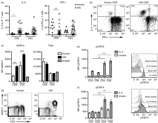
Differentiation of CD4(+) T cells into type 1 or type 2 subsets is mediated by the expression of the opposing lineage defining transcription factors T-bet and GATA-3. However, the existence of GATA-3(+) T-bet(+) CD4(+) T cells in mice suggests functional plasticity of these subsets. Little is known about type 1 and type 2 plasticity of human T-cell subsets in vivo. Here, we show that in the xenogeneic environment of humanized mice, which lacks a functional immune-regulatory network, human CD4(+) and, notably, CD8(+) T cells preferentially differentiate into interleukin (IL)-4(+) GATA-3(+) and IL-4(+) interferon-γ(+) GATA-3(+) T-bet(+) subsets. Treatment with recombinant human IL-12 or expansion of IL-12-producing human dendritic cells in vivo reverted this phenotype and led to the down-regulation of GATA-3 expression. These changes also correlated with improved antiviral immune responses in humanized mice. In conclusion, our study shows the capacity of human CD4(+) and CD8(+) T cells for stable co-expression of GATA-3 and T-bet in humanized mice and reveals a critical role for IL-12 in regulating this phenotype.

Keywords: T-cell plasticity; humanized mice; interleukin-12.


Figures

Figure 1
Figure 1
Preferential type 2 differentiation of human CD4+ and CD8+ T cells in humanized mice. HIS (human immune system) mice were generated by transplantation of human fetal liver-derived CD34+ haematopoietic stem cells into newborn NRG mice. Twelve weeks post transplantation type 1 versus type 2 differentiation of human CD3+ CD4+ and CD3+ CD8+ T cells present in the peripheral blood of HIS mice was compared with that of human peripheral blood mononuclear cell (PBMC) -derived T cells. (a) Intracellular interferon-γ (IFN-γ) and interleukin-4 (IL-4) production of human PBMC (n = 8) and HIS (n = 20) derived CD4+ and CD8+ T cells upon unspecific stimulation with PMA/ionomycin. (b) Representative FACS plots showing IFN-γ and IL-4 production of human and HIS CD8+ T cells. (c) Expression levels (mean fluorescence intensity) of the transcription factors GATA-3 and T-bet in CD4+ and CD8+ T cells isolated from human (n = 8) or HIS (n = 15) peripheral blood. (d) Representative FACS plots showing the expression of GATA-3 in human and HIS mouse-derived CD3+ T cells. (e,f) signal transducer and activator of transcription 6 (STAT-6) (e) and STAT-5 (f) phosphorylation of human (n = 8) and HIS mouse (n = 10) derived CD4+ and CD8+ T cells after 15 min of stimulation with human IL-4 and IL-2, respectively. Group data are shown on the left and representative histograms on the right. Data represent mean ± SEM. Summarized data of n = 8 human donors and n = 10 to n = 20 HIS mice acquired in at least three independent experiments are shown. Unpaired Student's t-test: *P ≤ 0·05; **P ≤ 0·005; ***P ≤ 0·0002.
Figure 2
Figure 2
High GATA-3 expression is specific for human T cells in humanized mice. (a) Frequencies of GATA-3+ human CD4+ and CD8+ T cells in human peripheral blood mononuclear cell (PBMC) (n = 8), human cord blood (n = 2), peripheral blood of HIS (human immune system) mice (n = 15) and two HLA-expressing humanized mouse models, HIS-HLA-A2 (n = 10) and BLT (bone marrow/liver/thymus) (n = 7). (b) Frequency of murine GATA-3+ CD4+ and CD8+ T cells derived from C57BL/6 mice (n = 5) and C57BL/6-NRG bone marrow chimeras (n = 5). (c) Representative FACS plots showing GATA-3 expression in human CD8+ T cells derived from human PBMC, human cord blood, HIS mice, HIS-HLA-A2 mice or BLT mice and in murine CD8+ T cells derived from C57BL/6 mice and C57BL/6-NRG bone marrow chimeras. Data represent mean ± SEM.
Figure 3
Figure 3
Co-expression of GATA-3 and T-bet in human CD4+ and CD8+ T cells in humanized mice. Twelve weeks post human CD34+ haematopoietic stem cell transplantation human naive CD45RA+ and effector CD45RO+ CD4+ and CD8+ T cells present in the blood of HIS (human immune system) mice were analysed for the expression of GATA-3 and T-bet by flow cytometry. (a) Representative FACS plots comparing the expression patterns of GATA-3 and T-bet in CD4+ and CD8+ T cells of human peripheral blood mononuclear cells (PBMC) and HIS mouse peripheral blood. (b) Representative FACS plots showing CD45RO expression on human and HIS CD8+ T cells. (c) Human and HIS CD8+ T cells were gated on CD45RO+ and CD45RA+ cells and T-bet and GATA-3 expression in these subsets was analysed. Representative histograms are shown. (d, e) Group data showing the frequencies of GATA-3 T-bet, GATA-3+ T-bet, GATA-3+ T-bet+, GATA-3 T-bet+ subsets within the CD45RO+ (left panel) and the CD45RA+ (right panel) populations of HIS mouse CD4+ (d) and CD8+ (e) T cells. Summarized data of n = 20 HIS mice acquired in three independent experiments are shown.
Figure 4
Figure 4
Co-expression profile of lineage transcription factors and cytokines in T cells of humanized mice. HIS (human immune system) mouse-derived peripheral CD4+ and CD8+ T cells (12 weeks post human CD34+ haematopoietic stem cells) were simultaneously analysed for the expression of the transcription factors GATA-3, T-bet, Foxp3 and Eomes and the production of the cytokines interferon (IFN-γ) and interleukin-4 (IL-4). (a) Representative FACS plots showing Foxp3 (upper left) and Eomes (upper right) expression within the CD3+ T-cell population of HIS mice. Cells gated on Foxp3+ and Foxp3 CD4+ T cells (lower left) and Eomes+ and Eomes CD8+ T cells (lower right) were then compared for co-expression of T-bet and GATA-3. (b) GATA-3 and T-bet co-expression patterns of CD4+ Foxp3+ (left) and CD8+ Eomes+ (right) T cells (n = 8). (c) Cells were stimulated ex vivo with PMA/ionomycin before intracellular staining for GATA-3, T-bet, IFN-γ and IL-4. Representative FACS plots show the expression of IFN-γ and IL-4 in CD8+ (upper panel) or CD4+ (lower panel) GATA-3+ T-bet and GATA 3+T-bet+ subsets. (d) Frequencies of IFN-γ+, IL-4+ and IFN-γ+/IL-4+ cells within the GATA-3+ T-bet and GATA-3+ T-bet+ subsets of HIS CD4+ and CD8+ T cells (n = 15). Data represent mean ± SEM. Summarized data of n = 8 to n = 15 HIS mice acquired in at least two independent experiments are shown. Unpaired Student's t-test: *P ≤ 0·05; ***P ≤ 0·0002.
Figure 5
Figure 5
Co-expression of GATA-3 and T-bet in single human CD4+ and CD8+ T cells is stable in vitro. (a) Concentrations of human and murine interleukin-12p70 (IL-12p70) and IL-4 and human interferon-γ (IFN-γ) in the serum of NRG mice (n = 10) and HIS (human immune system) mice (n = 18). (b, c) Purified HIS mouse-derived T cells were stimulated with anti-CD3/CD28-coated beads and cultured in vitro for 14 days without the addition of exogenous cytokines or in the presence of 10 ng/ml human recombinant IL-12p70. (b) Representative FACS plots show GATA-3 and T-bet expression patterns of purified CD4+ and CD8+ T cells directly post purification and at day 14 of in vitro culture. (c) Left: representative histograms show differences in GATA-3 expression levels of CD8+ T cells cultured ± human IL-12p70 for 14 days (−IL-12: grey solid; +IL-12 black open). Right: expression levels of GATA-3 in CD8+ and CD4+ cultured ± human IL-12p70 for 14 days. (d) Concentrations of human IL-4 and IFN-γ in supernatants of cultured T cells. Data represent mean ± SEM. Representative data of three independent in vitro experiments are shown. Unpaired Student's t-test: *P ≤ 0·05.
Figure 6
Figure 6
In vivo interleukin-12 (IL-12) treatment of humanized mice induces GATA-3 down-regulation and T-bet up-regulation in CD4+ and CD8+ T cells. HIS mice (12 weeks post human CD34+ haematopoietic stem cells) were injected intraperitoneally with three doses (day 0, 5, 8) of 1 μg/mouse recombinant human IL-12p70. Peripheral T cells were analysed directly pre IL-12 treatment and at day 12 post start of treatment. (a) T-bet (left graph) and GATA-3 (right graph) expression levels (mean fluorescence intensity, MFI) in CD8+ and CD4+ T cells pre and post IL-12 treatment (n = 13). (b, c) Frequencies of GATA-3 T-bet, GATA-3+ T-bet, GATA-3+ T-bet+, GATA-3 T-bet+ subsets within the CD45RO+ (left panel) and the CD45RA+ (right panel) populations of HIS CD8+ (b) and CD4+ (c) T cells pre and post IL-12 treatment (n = 9). (d) For the analysis of T-bet (left graph) and GATA-3 (right graph) expression in liver and spleen, intrahepatic and splenic T cells of mock (PBS) -treated mice (n = 12) were compared with IL-12-treated mice (day 12 post start of treatment) (n = 13) (e) Representative FACS plots showing intrahepatic GATA-3 T-bet, GATA-3+ T-bet, GATA-3+ T-bet+, GATA-3 T-bet+ CD4+ and CD8+ T-cell subsets in IL-12-treated and mock-treated mice. Data represent mean ± SEM. Summarized data of four independent experiments are shown. Unpaired Student's t-test: *P ≤ 0·05; **P ≤ 0·005; ***P ≤ 0·0002.
Figure 7
Figure 7
Interleukin-12 (IL-12) -induced change of GATA-3 and T-bet expression patterns is not dependent on T-cell proliferation and is linked to increased interferon-γ (IFN-γ) production. (a, b) Cells of IL-12-treated HIS (human immune system) mice were stained pre and post treatment for intracellular expression of GATA-3, T-bet and ki67 to track the proliferation of T-cell subsets in vivo. (a) Frequencies of ki67+ CD8+ and CD4+ T cells in HIS mice pre and post IL-12 treatment (n = 9). (b) Representative FACS plots showing ki67 expression in CD8+ T cells from one HIS mouse pre and post IL-12 treatment and representative FACS plots showing GATA-3 and T-bet expression in gated ki67 and ki67+ CD8+ T cells pre and post IL-12 treatment. (c) Blood-derived cells of IL-12-treated HIS mice (n = 13) and controls (n = 20) were stimulated ex vivo with PMA/ionomycin followed by intracellular staining for IL-4 and IFN-γ. Frequencies of IFN-γ (upper graph), IL-4 (middle graph) and IFN-γ/IL-4 (lower graph) producing CD4+ and CD8+ T cells are shown. Summarized data of four independent experiments are shown. Unpaired Student's t-test: **P ≤ 0·005; ***P ≤ 0·0002.
Figure 8
Figure 8
Flt3L-induced in vivo expansion of dendritic cells (DC) results in GATA-3 down-regulation in CD4+ and CD8+ T cells. HIS (human immune system) mice (12 weeks post human CD34+ HSC) were treated with recombinant human Flt3L (10 μg/mouse intraperitoneal) for 8 consecutive days. At day 10 post stop of treatment mice were analysed for human DC subsets, cytokine serum concentrations and GATA-3 and T-bet expression in T cells. (a) Frequencies of BDCA-1+, BDCA-2+, BDCA-3+ DC subsets in the blood of Flt3L-treated mice and controls (n = 9). (b) Representative FACS plots showing BDCA-3 and BDCA-1 DCs in Flt3l-treated mice and controls (PBS). (c) Serum concentrations of human IL-4 and human IL-12 in Flt3L-treated mice and controls (n = 9). (d) GATA-3 (left graph) and T-bet (right graph) expression levels (mean fluorescence intensity) in peripheral HIS CD8+ and CD4+ T cells pre and post Flt3L treatment (n = 9). (e, f) Frequencies of GATA-3 T-bet, GATA-3+ T-bet, GATA-3+ T-bet+, GATA-3 T-bet+ subsets within the CD8+ (e) and CD4+ (f) T-cell population pre and post Flt3L treatment (n = 9). Data represent mean ± SEM. Summarized data of two independent experiments are shown. Unpaired Student's t-test: *P ≤ 0·05; **P ≤ 0·005; ***P ≤ 0·0002.
Figure 9
Figure 9
Antiviral immunity is enhanced in interleukin-2 (IL-12) -treated humanized mice. (a) HIS (human immune system) mice were infected intravenously with 5 × 10⁹ particles of a hepatotropic replication-deficient E1/E2-deleted adenovirus serotype 5 expressing firefly luciferase (AdV5-Fluc) and treated with recombinant human IL-12 (1 μg/mouse) as shown in the schematic. (b) Fluc-expression was longitudinally monitored in IL-12-treated mice (+IL-12) and controls (−IL-12) using an in vivo imaging system (IVIS) and quantified in photons/second. Left: A representative picture shows Fluc-expression in IL-12-treated mice (+IL-12) and controls (−IL-12) at day 20 post infection. Right: Group data showing Fluc-expression in IL-12 treated mice (+IL-12) and controls (−IL-12) at day 20 post infection (n = 10). (c) Frequencies of GATA-3 T-bet, GATA-3+ T-bet, GATA-3+ T-bet+, GATA-3 T-bet+ CD4+ (upper graph) and CD8+ (lower graph) T-cell subsets in the blood of HIS mice pre AdV5 infection and at day 20 post infection (±IL12) (n = 6). (d) Representative FACS plots showing GATA-3 and T-bet expression patterns in intrahepatic CD8+ T cells of AdV5-infected HIS mice ±IL12 treatment at day 20 post infection. (e) Serum concentrations of human IFN-γ and IL-4 in AdV5-infected mice ± IL12 treatment and controls (n = 6). Summarized data of at least two independent experiments are shown. Data represent mean ± SEM. Unpaired Student's t-test: *P ≤ 0·05; **P ≤ 0·005.

References

    1. Zhu J, Yamane H, Paul WE. Differentiation of effector CD4 T cell populations (*) Annu Rev Immunol. 2010;28:445–89. - PMC - PubMed
    1. Amsen D, Spilianakis CG, Flavell RA. How are TH1 and TH2 effector cells made? Curr Opin Immunol. 2009;21:153–60. - PMC - PubMed
    1. Mosmann TR, Coffman RL. TH1 and TH2 cells: different patterns of lymphokine secretion lead to different functional properties. Annu Rev Immunol. 1989;7:145–73. - PubMed
    1. Pulendran B, Artis D. New paradigms in type 2 immunity. Science. 2012;337:431–5. - PMC - PubMed
    1. Szabo SJ, Kim ST, Costa GL, Zhang X, Fathman CG, Glimcher LH. A novel transcription factor, T-bet, directs Th1 lineage commitment. Cell. 2000;100:655–69. - PubMed

Publication types

MeSH terms

LinkOut - more resources