Comparative Study
. 2014 Oct;143(2):258-68.
doi: 10.1111/imm.12306.

Mycobacterium indicus pranii and Mycobacterium bovis BCG lead to differential macrophage activation in Toll-like receptor-dependent manner

Affiliations
Comparative Study

Mycobacterium indicus pranii and Mycobacterium bovis BCG lead to differential macrophage activation in Toll-like receptor-dependent manner

Pawan Kumar et al. Immunology. 2014 Oct.

Abstract

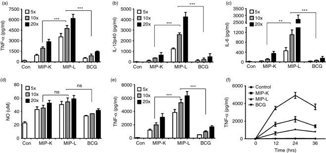
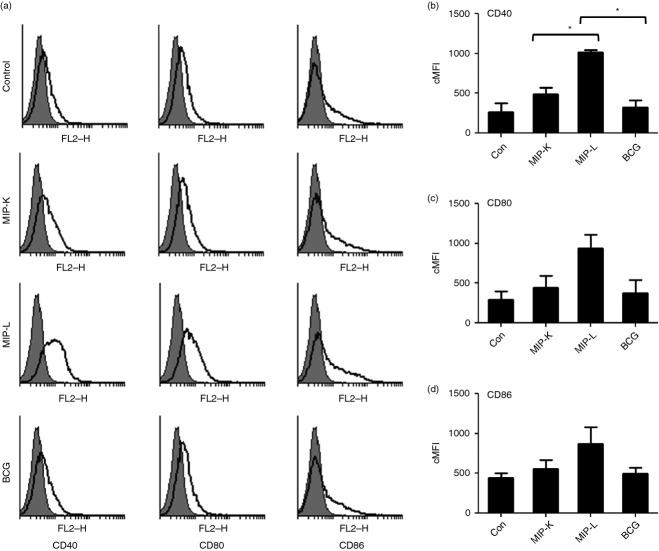
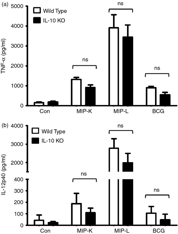
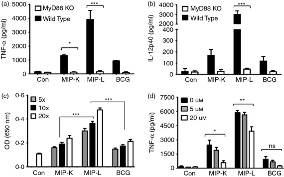
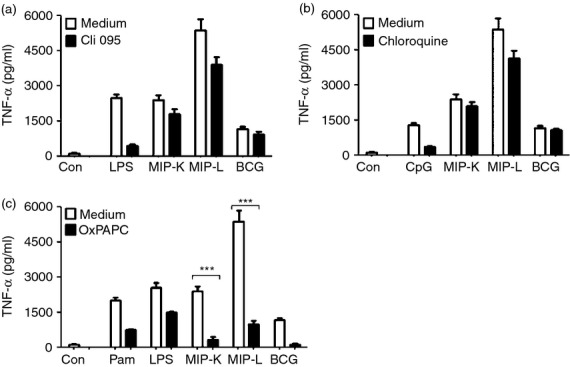
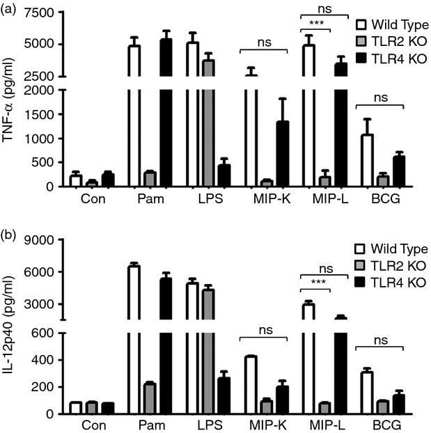
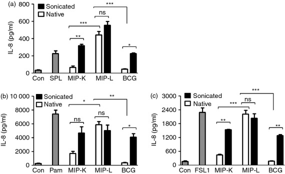
Mycobacterium indicus pranii (MIP) is an atypical mycobacterial species possessing strong immunomodulatory properties. It is a potent vaccine candidate against tuberculosis, promotes Th1 immune response and protects mice from tumours. In previous studies, we demonstrated higher protective efficacy of MIP against experimental tuberculosis as compared with bacillus Calmette-Guérin (BCG). Since macrophages play an important role in the pathology of mycobacterial diseases and cancer, in the present study, we evaluated the MIP in live and killed form for macrophage activation potential, compared it with BCG and investigated the underlying mechanisms. High levels of tumour necrosis factor-α, interleukin-12p40 (IL-12p40), IL-6 and nitric oxide were produced by MIP-stimulated macrophages as compared with BCG-stimulated macrophages. Prominent up-regulation of co-stimulatory molecules CD40, CD80 and CD86 was also observed in response to MIP. Loss of response in MyD88-deficient macrophages showed that both MIP and BCG activate the macrophages in a MyD88-dependent manner. MyD88 signalling pathway culminates in nuclear factor-κB/activator protein-1 (NF-κB/AP-1) activation and higher activation of NF-κB/AP-1 was observed in response to MIP. With the help of pharmacological inhibitors and Toll-like receptor (TLR) -deficient macrophages, we observed the role of TLR2, TLR4 and intracellular TLRs in MIP-mediated macrophage activation. Stimulation of HEK293 cells expressing TLR2 in homodimeric or heterodimeric form showed that MIP has a distinctly higher level of TLR2 agonist activity compared with BCG. Further experiments suggested that TLR2 ligands are well exposed in MIP whereas they are obscured in BCG. Our findings establish the higher macrophage activation potential of MIP compared with BCG and delineate the underlying mechanism.

Keywords: Toll-like receptors; bacterial; cytokines.


Figures

Figure 1
Figure 1
Production of pro-inflammatory cytokines and nitric oxide (NO) by heat-killed Mycobacterium indicus pranii (MIP-K), live Mycobacterium indicus pranii (MIP-L) and bacillus Calmette–Guérin (BCG) -stimulated macrophages. Peritoneal macrophages were stimulated with MIP-K, MIP-L and BCG and culture supernatants were analysed by ELISA. Higher levels of tumour necrosis factor-α (TNF-α), interleukin-12p40 (IL-12p40) and IL-6 production was observed in response to MIP-L. MIP-K led to moderate level of these cytokines (a–c). High level of NO was also observed in MIP-L and MIP-K-stimulated macrophages compared with BCG, but difference was not significant. (d). RAW 264.7 macrophages were also stimulated with MIP-K, MIP-L and BCG, and a high level of TNF-α was produced in response to MIP-L. A moderate level of TNF-α was produced by MIP-K-stimulated RAW 264.7 macrophages (e). Time-kinetics of RAW 264.7 macrophage response to MIP-K, MIP-L and BCC was studied and at all time-points, higher levels of TNF-α production were observed in response to MIP-L followed by MIP-K (f). Data shown are mean ± SEM of three independent experiments. **P < 0·01, ***P < 0·001 and ns, not significant.
Figure 2
Figure 2
Expression of activation markers by macrophages stimulated with heat-killed Mycobacterium indicus pranii (MIP-K), live Mycobacterium indicus pranii (MIP-L) and bacillus Calmette–Guérin (BCG). RAW 264.7 macrophages were stimulated with MIP-K, MIP-L and BCG at a multiplicity of stimulation of 10. Cells were stained with phycoerythrin-conjugated anti-mouse CD40, CD80 and CD86 monoclonal antibodies and analysed by flow cytometry. MIP-L was leading to higher up-regulation of co-stimulatory molecules. Moderate up-regulation was observed in response to MIP-K. Representative histograms are shown. Composite Mean Fluorescence Intensity (= % positive cells × mean fluorescence intensity of positive cells) is also shown as a bar graph. Data shown are mean ± SEM of two independent experiments. *P < 0·05.
Figure 3
Figure 3
Response of interleukin-10 deficient (IL-10/) and wild-type macrophages to heat-killed Mycobacterium indicus pranii (MIP-K), live Mycobacterium indicus pranii (MIP-L) and bacillus Calmette–Guérin (BCG). Peritoneal macrophages from IL-10/ and wild-type mice were stimulated with MIP-K, MIP-L and BCG at a multiplicity of stimulation of 10. A similar response in terms of tumour necrosis factor-α (TNF-α) and IL-12-p40 production was observed in IL-10/ and wild-type macrophages. Data shown are mean ± SEM of three independent experiments. ns, not significant.
Figure 4
Figure 4
Role of MyD88 and nuclear factor-κB/activator protein 1 ((NF-κB/AP-1) in heat-killed Mycobacterium indicus pranii (MIP-K), live Mycobacterium indicus pranii (MIP-L) and bacillus Calmette–Guérin (BCG) mediated activation of macrophages. Peritoneal macrophages from MyD88/ and wild-type mice were stimulated with MIP-K, MIP-L and BCG. Response to MIP and BCG was lost in MyD88/ macrophages, as shown by drastically reduced levels of tumour necrosis factor-α (TNF-α) and interleukin-12 p40 (IL-12p40) in culture supernatants (a,b). Role of NF-κB/AP-1 in macrophage activation by MIP or BCG was determined by stimulating RAW-Blue cells, which express NF-κB/AP-1 inducible secreted alkaline phosphatase (SEAP). Higher levels of SEAP activity were observed in response to MIP-L (c). The role of NF-κB/AP-1 in MIP-mediated macrophage activation was determined with the help of pharmacological inhibitor. RAW 264.7 macrophages were incubated with curcumin at the indicated concentration for 30 min and then stimulated with MIP-K, MIP-L and BCG. Dose-dependent decrease in TNF-α production in response to MIP-K, MIP-L and BCG was observed in the presence of curcumin (d). Data shown are mean ± SEM of three independent experiments. *P < 0·05, **P < 0·01, ***P < 0·001 and ns, not significant.
Figure 5
Figure 5
Role of Toll-like receptors (TLRs) in macrophage activation by heat-killed Mycobacterium indicus pranii (MIP-K), live Mycobacterium indicus pranii (MIP-L) and bacillus Calmette–Guérin (BCG). RAW 264.7 macrophages were incubated with 1·5 μg/ml Cli095 (TLR4 inhibitor), 80 μg/ml OxPAPC (TLR2 and TLR4 inhibitor) or 20 μm chloroquine (intracellular TLR inhibitor) for 60 min. MIP-K, MIP-L and BCG were subsequently added at multiplicities of infection of 10 and culture supernatants were analysed for tumour necrosis factor-α (TNF-α) after 24 hr. Diminished levels of TNF-α production by MIP-K, MIP-L and BCG stimulated macropahges were observed in the presence of Cli095 (a). Decreased TNF-α production by MIP-L-stimulated macrophages was also observed in the presence of chloroquine (b). Dramatic reduction in TNF-α production by MIP-K, MIP-L and BCG-stimulated macrophages was observed in the presence of OxPAPC (c). Pam-Pam3CSK4, LPS-lipopolysachharide, CpG-CpG DNA were used as positive controls. Data shown are mean ± SEM of three independent experiments. ***P < 0·001 and ns, not significant.
Figure 6
Figure 6
Response of Toll-like receptor (TLR) -deficient macrophages to heat-killed Mycobacterium indicus pranii (MIP-K), live Mycobacterium indicus pranii (MIP-L) and bacillus Calmette–Guérin (BCG). Peritoneal macrophages from TLR2/, TLR4/ and wild-type mice were stimulated with MIP-K, MIP-L and BCG at multiplicities of infection of 10. Culture supernatants were analysed for tumour necrosis factor-α (TNF-α) and interleukin-12 p40 (IL-12p40) by ELISA. Diminished production of TNF-α and IL-12p40 in response to MIP-K, MIP-L and BCG was observed in TLR4/ macrophages. Dramatic reduction in the TNF-α and IL-12p40 production in response to MIP-K, MIP-L and BCG was observed in TLR2/ macrophages. Pam (Pam3CSK4, 10 ng/ml) and LPS (Lipopolysaccharide, 10 ng/ml) were used as positive controls. Data shown are mean ± SEM of two independent experiments. ***P < 0·001 and ns, not significant.
Figure 7
Figure 7
Delineation of Toll-like receptor (TLR2) signalling by heat-killed Mycobacterium indicus pranii (MIP-K), live Mycobacterium indicus pranii (MIP-L) and bacillus Calmette–Guérin (BCG). HEK293 cells expressing TLR2/2, TLR2/1 or TLR2/6 (a, b and c, respectively) were stimulated with MIP-K, MIP-L and BCG in native or sonicated form at a multiplicity of infection of 50. Culture supernatants were collected after 48 hr and analysed for interleukin-8 (IL-8) by ELISA. Higher levels of IL-8 production from 293tlr2/2, 293tlr2/1 and 293tlr2/6 cells were observed in response to native and sonicated MIP-L. Maximum level of IL-8 was produced by 293tlr2/1 cells. Native MIP-K led to moderate levels of IL-8 from 293tlr2/2, 293tlr2/1 and 293tlr2/6 cells. Native BCG did not lead to IL-8 production from either of 293tlr2/2, 293tlr2/1 and 293tlr2/6 cells, indicating that TLR2 ligands are not well exposed in native BCG. Substantial levels of IL-8 were produced by 293tlr2/2, 293tlr2/1 and 293tlr2/6 cells in response to sonicated BCG. SPL-Streptococcus pneumoniae lysate (TLR2/2 ligand, MOS = 50), Pam-Pam3CSK4 (TLR2/1 ligand, 0·2 μg/ml), FSL1 (TLR2/6 ligand, 1 μg/ml) were used as positive controls. Data shown are mean ± SEM of three independent experiments. *P < 0·05, **P < 0·01, ***P < 0·001 and ns, not significant.

References

    1. Saxena VK, Singh US, Singh AK. Bacteriological study of a rapidly growing strain of Mycobacterium. Lepr India. 1978;50:588–96. - PubMed
    1. Deo MG. Anti-leprosy vaccines – field trials and future prospects. Indian J Lepr. 1984;56:764–75. - PubMed
    1. Talwar GP, Zaheer SA, Mukherjee R, et al. Immunotherapeutic effects of a vaccine based on a saprophytic cultivable mycobacterium, Mycobacterium w in multibacillary leprosy patients. Vaccine. 1990;8:121–9. - PubMed
    1. Katoch K, Katoch VM, Natrajan M, et al. Treatment of bacilliferous BL/LL cases with combined chemotherapy and immunotherapy. Int J Lepr Other Mycobact Dis. 1995;63:202–12. - PubMed
    1. Sharma P, Misra RS, Kar HK, Mukherjee A, Poricha D, Kaur H, Mukherjee R, Rani R. Mycobacterium w vaccine, a useful adjuvant to multidrug therapy in multibacillary leprosy: a report on hospital based immunotherapeutic clinical trials with a follow-up of 1–7 years after treatment. Lepr Rev. 2000;71:179–92. - PubMed

Publication types

MeSH terms

LinkOut - more resources