. 2014 Apr 24;157(3):689-701.
doi: 10.1016/j.cell.2014.02.024.

Identification of a circadian output circuit for rest:activity rhythms in Drosophila

Affiliations

Identification of a circadian output circuit for rest:activity rhythms in Drosophila

Daniel J Cavanaugh et al. Cell. .

Abstract

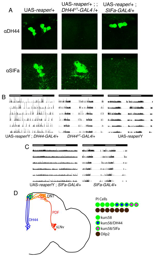
Though much is known about the cellular and molecular components of the circadian clock, output pathways that couple clock cells to overt behaviors have not been identified. We conducted a screen for circadian-relevant neurons in the Drosophila brain and report here that cells of the pars intercerebralis (PI), a functional homolog of the mammalian hypothalamus, comprise an important component of the circadian output pathway for rest:activity rhythms. GFP reconstitution across synaptic partners (GRASP) analysis demonstrates that PI cells are connected to the clock through a polysynaptic circuit extending from pacemaker cells to PI neurons. Molecular profiling of relevant PI cells identified the corticotropin-releasing factor (CRF) homolog, DH44, as a circadian output molecule that is specifically expressed by PI neurons and is required for normal rest:activity rhythms. Notably, selective activation or ablation of just six DH44+ PI cells causes arrhythmicity. These findings delineate a circuit through which clock cells can modulate locomotor rhythms.


Figures

Figure 1
Figure 1
Constitutive activation of non-Dilp2-expressing PI neurons causes behavioral arrhythmicity without affecting the molecular clock. (A) Maximum projection confocal brain images of GAL4 hits. GAL4 hits were crossed to UAS-nlsGFP flies, and brains were stained for GFP. Insets show close-up of PI region. (B) Representative activity records of individual kurs58-GAL4/UAS-dTrpA1 (left), kurs58-GAL4/+ (middle), and UAS-dTrpA1/+ flies under DD conditions, before and after transition to 28°C. Activity records are double-plotted, with gray and black bars indicating subjective day and night, respectively, for the last 4 days at 21°C, and the first 4 days at 28°C (red shading). (C) kurs58-GAL4/UAS-nlsGFP ; Dilp2-mCherry/+ brain stained for GFP (right; green) and mCherry (left; red). A merged image demonstrating lack of overlap is shown in the center panel. (D) Activity records of individual Dilp2-GAL4/UAS-dTrpA1 (left), Dilp2-GAL4/+ (middle), and UAS-dTrpA1/+ flies under DD conditions before and after transition to 28°C. (E) kurs58-GAL4/UAS-dTrpA1 and (F) control dTrpA1/+ brains were stained for PERIOD (red) and PDF (blue) at various time points on the second day of DD at 28°C. PER cycling was indistinguishable between the two genotypes. (G) Activity records of individual kurs58-GAL4/UAS-dTrpA1 (left), kurs58-GAL4/+ (middle) and UAS-dTrpA1/+ (right) flies in DD conditions for 5 days at 21°C, followed by 4 days at 28°C (red shading), and 5 days at 21°C, demonstrating recovery of rhythms after transition back to 21°C. See also Figure S1.
Figure 2
Figure 2
GRASP analysis reveals a circadian output circuit emanating from clock neurons. (A) Pdf-LexA,LexAop-GFP11/LexAop-FLP ; nsyb-GAL4/FRT-UAS-GFP1–10-FRT brain visualized for GFP (green). PDF staining (blue) shows that GRASP signal is visible along the length of the dorsal projection of the s-LNvs. (B) cry-GAL4/Y ; Pdf-LexA,LexAop-GFP11/Pdf-GAL80 ; UAS-GFP1–10/+ brain visualized for GFP (green) and PDF (blue), with identical GRASP signal as in (A). (C) Pdf-LexA,LexAop-GFP11/+ ; Clk4.1M-GAL4/UAS-GFP1–10 brain showing GRASP signal in dorsal brain. (D) Close-up of boxed region in C, double-labeled for PER immunofluorescence (red). (E) TUG-GAL4/UAS-nlsGFP ; 911-QF/QUAS-mtd-tomato brain stained for tomato. (F) Close-up of boxed region in (E), double-labeled for GFP, showing that 911-QF expresses in DN1 cells. (G) Clk4.1-LexA/LexAop-CD8GFP brain stained for GFP, showing specific expression in DN1 cells. (H) Clk4.1-LexA/LexAop-CD8GFP brain stained for GFP (green) and PER (red). (I) kurs58-GAL4/LexAop-GFP11 ; Clk4.1-LexA/UAS-GFP1–10 brain, with GRASP signal (green) in PI region (boxed). Brain is co-labeled for PDF (blue). (J) Close-up of boxed region in (I). (K) kurs58-GAL4/QUAS-GFP11 ; 911-QF/UAS-GFP1–10 brain confirms GRASP signal in PI region. (L) kurs58-GAL4/QUAS-Rab3eGFP ; 911-QF/UAS-Denmark brain, double labeled for GFP (green) and mCherry (red). For panels (A) and (B), dotted line indicates brain surface. All GRASP signals represent endogenous GFP fluorescence (no GFP antibody was used). See also Figure S2.
Figure 3
Figure 3
DH44 is a circadian signaling molecule expressed by PI cells. (A) DH44 immunostaining. (B) Close-up of boxed region in (A), showing expression in 6 PI neurons. (C) Overlap between DH44 immunostaining (blue) and kurs58-GAL4>nlsGFP (green). (D) Dilp2-mCherry brain, double-labeled for mCherry (green) and DH44 (blue), showing lack of overlap. (E–F) Partial reduction of DH44 immunostaining in kurs58-GAL4/UAS-DH44 RNAi KK ; UAS-Dicer2/+ and kurs58-GAL4/+ ; UAS-DH44 RNAi TRiP/UAS-Dicer2 brains. (G–H) Complete loss of DH44 immunostaining in elav-GAL4/Y ; UAS-DH44 RNAi KK/UAS-Dicer2 or elav-GAL4/Y ; UAS-Dicer2/+ ; UAS-DH44 RNAi TRiP/+ brain. (I–J) Individual activity records demonstrating weakened rhythms following pan-neuronal knockdown of DH44 in elav-GAL4/Y ; UAS-DH44 RNAi KK/UAS-Dicer2 flies and elav-GAL4/Y ; UAS-Dicer2/+ ; UAS-DH44 RNAi TRiP/+ flies. (K) Tub-GAL4/+ ; UAS-DH44 Antagonist/+ flies demonstrate that DH44 receptor antagonism degrades rest:activity rhythms. See also Figure S3.
Figure 4
Figure 4
Activation of DH44+ PI neurons degrades rest:activity rhythms. (A) Schematic of the DH44 gene, showing the location of the regulatory sequences used to drive GAL4 expression for the DH44-GAL4 lines. (B) DH44VT-GAL4/UAS-CD8GFP brain, stained for GFP. (C–E) UAS-nlsGFP/+ ; DH44VT-GAL4/+ brain, double-labeled for GFP (C) and DH44 (E) immunofluorescence. A merged image is shown in (D), demonstrating complete overlap. (F) DH44FL-GAL4/UAS-CD8GFP brain, stained for GFP. (G) Activity records of individual UAS-dTrpA1/+ ; DH44VT-GAL4/+ (left), DH44VT-GAL4/+ (middle), and UAS-dTrpA1/+ (right) flies under DD conditions before and after transition to 28°C (red shading). (H) Activity records of individual UAS-dTrpA1/+ ; DH44-GAL4FL/+ (left) and DH44FL-GAL4/+ (right) under DD conditions before and after transition to 28°C (red shading).
Figure 5
Figure 5
Ablation of subsets of PI neurons results in behavioral arrhythmicity. (A) Complete and specific ablation of the DH44+ or SIFa+ subsets of PI neurons in UAS-reaper/+ ; ; DH44VT-GAL4/+ and UAS-reaper/+ ; SIFa-GAL4/+ flies, respectively. Ablation is demonstrated by a loss of DH44 (top panels) or SIFa (bottom panels) immunofluorescence. (B) Activity plots of individual UAS-reaper/Y ; ; DH44VT-GAL4/+ (left), DH44VT-GAL4/+ (middle) and UAS-reaper/Y (right) flies in DD conditions. (C) Activity plots of individual UAS-reaper/Y ; SIFa-GAL4/+ (left) and SIFa-GAL4/+ (right) flies in DD conditions. (D) Model of the circadian output circuit for locomotor rhythms. One hemisphere of the fly brain is depicted. The circuit extends from the master pacemaker s-LNvs (red), through DN1s (orange), and onto kurs58+, DH44+ PI cells (blue), which modulate locomotor rhythms through the release of DH44, in addition to other, unknown factors. The s-LNvs likely control DN1 cells through the release of PDF. PI cells can be divided into Dilp2+ cells (7/hemisphere; brown) and kurs58-GAL4+ cells (~9/hemisphere; green). kurs58-GAL4+ cells can be further subdivided into SIFa+ (2/hemisphere; pink with green outline), and DH44+ (3/hemisphere; blue with green outline). There are ~4 additional kurs58-GAL4+ cells that express neither DH44 nor SIFa. For simplicity only DH44+ and SIFa+ PI cells are depicted in the brain schematic. See also Figures S4.

Similar articles

Cited by

References

    1. Allada R, Chung BY. Circadian organization of behavior and physiology in Drosophila. Annual Rev of Physiol. 2009;72:605–624. - PMC - PubMed
    1. Balsalobre A, Brown SA, Marcacci L, Tronche F, Kellendonk C, Reichardt HM, Schütz G, Schibler U. Resetting of circadian time in peripheral tissues by glucocorticoid signaling. Science. 2000;289:2344–7. - PubMed
    1. Belgacem YH, Martin J-R. Neuroendocrine control of a sexually dimorphic behavior by a few neurons of the pars intercerebralis in Drosophila. Proc Natl Acad Sci USA. 2002;99:15154–58. - PMC - PubMed
    1. Broughton SJ, Piper MD, Ikeya T, Bass TM, Jacobson J, Driege Y, Martinez P, Hafen E, Withers DJ, Leevers SJ, et al. Longer lifespan, altered metabolism, and stress resistance in Drosophila from ablation of cells making insulin-like ligands. Proc Natl Acad Sci USA. 2005;102:3105–3110. - PMC - PubMed
    1. Buckley PT, Lee MT, Sul JY, Miyashiro KY, Bell TJ, Fisher SA, Kim J, Eberwine J. Cytoplasmic intron sequence-retaining transcripts can be dendritically targeted via ID element retrotransposons. Neuron. 2011;69:877–84. - PMC - PubMed

Publication types