

Stress, inflammation, and defense of homeostasis
- PMID: 24766892
- PMCID: PMC4048989
- DOI: 10.1016/j.molcel.2014.03.030
Stress, inflammation, and defense of homeostasis
Abstract
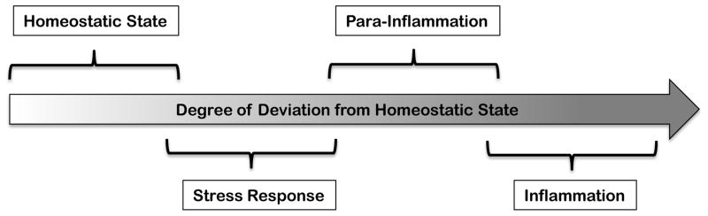
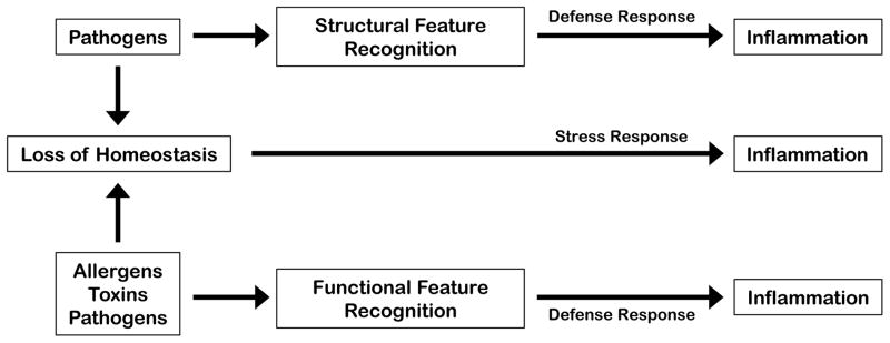
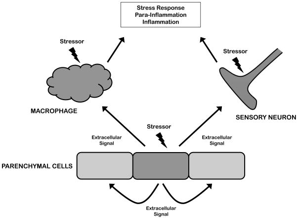
Inflammation is traditionally considered a defense response induced by infection or injury. However, inflammation can also be induced by tissue stress and malfunction in the absence of infection or overt tissue damage. Here we discuss the relationship between homeostasis, stress responses, and inflammation. Stress responses have cell-autonomous and cell-extrinsic components, the latter contributing to tissue level adaptation to stress conditions. Inflammation can be thought of as the extreme end of a spectrum that ranges from homeostasis to stress response to bona fide inflammatory response. Inflammation can be triggered by two types of stimuli: extreme deviations of homeostasis or challenges that cause a disruption of homeostasis. This perspective may help to explain qualitative differences and functional outcomes of diverse inflammatory responses.
Copyright © 2014 Elsevier Inc. All rights reserved.
Figures
References
-
- Allavena P, Sica A, Solinas G, Porta C, Mantovani A. The inflammatory micro-environment in tumor progression: the role of tumor-associated macrophages. Critical reviews in oncology/hematology. 2008;66:1–9. - PubMed
-
- Anckar J, Sistonen L. Heat shock factor 1 as a coordinator of stress and developmental pathways. Advances in experimental medicine and biology. 2007;594:78–88. - PubMed
-
- Aramburu J, Drews-Elger K, Estrada-Gelonch A, Minguillón J, Morancho B, Santiago V, López-Rodríguez C. Regulation of the hypertonic stress response and other cellular functions by the Rel-like transcription factor NFAT5. Biochemical pharmacology. 2006;72:1597–1604. - PubMed
Publication types
MeSH terms
Grants and funding
LinkOut - more resources
Full Text Sources
Other Literature Sources
