. 2014 May;39(5):233-44.
doi: 10.1016/j.tibs.2014.03.002. Epub 2014 Apr 21.

Allosteric sodium in class A GPCR signaling

Affiliations

Allosteric sodium in class A GPCR signaling

Vsevolod Katritch et al. Trends Biochem Sci. 2014 May.

Abstract

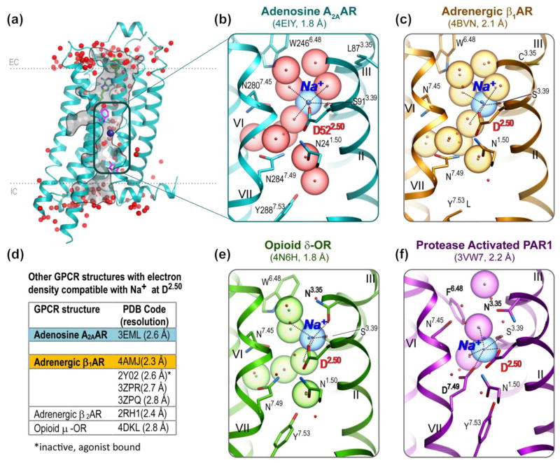
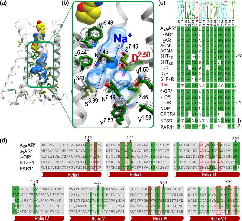
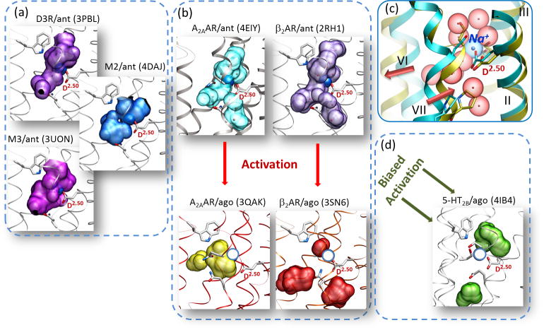
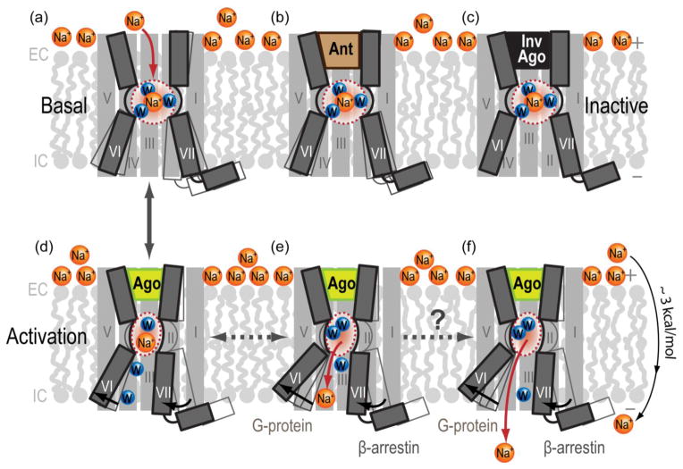
Despite their functional and structural diversity, G-protein-coupled receptors (GPCRs) share a common mechanism of signal transduction via conformational changes in the seven-transmembrane (7TM) helical domain. New major insights into this mechanism come from the recent crystallographic discoveries of a partially hydrated sodium ion that is specifically bound in the middle of the 7TM bundle of multiple class A GPCRs. This review discusses the remarkable structural conservation and distinct features of the Na(+) pocket in this most populous GPCR class, as well as the conformational collapse of the pocket upon receptor activation. New insights help to explain allosteric effects of sodium on GPCR agonist binding and activation, and sodium's role as a potential co-factor in class A GPCR function.

Keywords: GPCR activation; allosteric modulation; biased signaling; conserved pocket; sodium ion; water binding.


Figures

Figure 1
Figure 1
Na+ and water cluster detected in G protein-coupled receptor (GPCR) structures. (A) The high-resolution A2A adenosine receptor (A2AAR) structure [8] shown with waters (red spheres) and Na+ (blue sphere) in the narrow passage connecting the extracellular (EC) and intracellular (IC) sides of the receptor. (B) Close-up of the A2AAR allosteric pocket comprising Na+ and a cluster of 10 water molecules. Acidic residue D2.50 and all residue positions of the pocket involved in polar interaction with the cluster are shown as sticks. Roman numerals show numbering of the transmembrane helices. (C) Close-up of the β1AR allosteric pocket [13], colored orange. Ten water molecules and the Na+ position from A2AAR are shown for comparison as red dots and a blue dot, respectively. Note the Y3437.53L mutation. (D) A list of medium-resolution structures (2.3–2.8 Å) [,,–107] with electron densities in the proximity of D2.50 that are potentially compatible with sodium binding. (E) Close-up of the δ-opioid receptor (δ-OR) allosteric pocket [15], colored green. (F) Close-up of the protease-activated receptor (PAR1) allosteric pocket [14], colored magenta.
Figure 2
Figure 2
Structural and sequence conservation of the Na+ and water pocket in G protein-coupled receptors (GPCRs). (A) Overview of the A2A adenosine receptor (A2AAR) crystal structure, showing residues with higher than 50% conservation in all non-olfactory class A GPCRs as sticks with green carbons. (B) A close-up of the central allosteric pocket (transparent blue surface), showing the side chains located within 5 Å from the 10 waters of the sodium ion/water cluster (green sticks: A2AAR; gray thin lines: the corresponding side chains of the overlaid GPCR crystal structures in inactive state). The helix VII backbone in the foreground has been removed for clarity. (C) Sequence conservation of the 16 pocket residues. The top part shows the residue conservation profile in all non-olfactory class A GPCRs, where the height of the residue letter represents the share of the residue in this position. The bottom part shows individual residues in all available class A crystal structures, with conserved residues highlighted in green. Greek letters on the right denote the four major branches of class A GPCRs. Receptors with sodium binding determined by a high-resolution crystal structure are in bold and marked with ‘*’. Rhodopsin, which lacks a Na+ binding pocket, is shown in red. (D) Sequence alignment in seven-transmembrane helices of representative class A GPCRs from all four major branches. Residues of the Na+ pocket are highlighted by red boxes, and conserved residues are highlighted in green. The most conserved residues in each helix (X.50) are marked by arrows.
Figure 3
Figure 3
Activation involves collapse of the sodium pocket in class A G protein-coupled receptors (GPCRs). (A) Activation-related conformational changes, in particular inward movement of helix VII and outward movement of helix VI, cause collapse and relocation of the allosteric pocket in A2A adenosine receptor (A2AAR), which makes it incompatible with binding of the Na+ and water cluster. (B) Collapse of the pocket between inactive and activated states found in A2AAR structures (PDB identifiers 4EIY and 3QAK, respectively) is even more pronounced in β2-adrenergic receptors ((PDB identifiers 2RH1 and 3SN6). (C) Most other inactive class A GPCR structures have pockets that are compatible with sodium binding. (D) The pocket is also collapsed in the arrestin-biased activated structure of 5HT2B (right bottom panel). Allosteric pockets are shown by semitransparent surfaces, and the D2.50 side chain is labeled in all panels. The position of Na+ in the A2AAR-antagonist complex (PDB identifier 4EIY) superimposed on the other inactive structures is shown as dark blue spheres, whereas Na+ superimposed on the active-state structures is indicated as empty circles in light blue.
Figure 4
Figure 4
Hypothetical mechanisms of Na+ involvement in GPCR activation. (A) In the inactive apostate receptors, sodium gains access from the extracellular side into the conserved allosteric pocket, where it forms a network of ionic and polar interactions as a part of Na+ and water cluster. (B) Antagonist (Ant) or (C) inverse agonist (InvAgo) binding in the orthosteric pocket is compatible with allosteric Na+ binding and can further stabilize the inactive state. (D) GPCR activation by agonists (Ago), as seen in crystal structures of activated receptors, involves an inward movement of helix VII and an outward movement of helix VI, leading to a partial collapse/reshaping of the allosteric pocket. The resulting collapse of an optimal Na+/water cluster potentiates displacement of Na+ along the internal passage towards the intracellular side of membrane, where it can either (E) engage in transient interactions with other conserved acidic residues [e.g., D(E)3.49 of the D(E)RY motif] and G proteins or (F) exit the protein altogether, traversing the membrane along the Na+ electrochemical gradient, estimated at about 3 kcal/mol.
Figure 5
Figure 5
Predicted binding of amilorides in the conserved allosteric Na+ pocket. The flexible docking poses of amiloride (A) and 5-(N,N-Hexamethylene)amiloride (HMA) (B) in the sodium pocket of inactive A2AAR suggest that both ligands optimally fit the sodium cavity with only slight conformational changes from the crystal structure (PDB identifier: 4EIY). Neither amiloride ligand (shown with carbon atoms colored magenta) makes direct contact with the orthosteric antagonist ZM241385 (with yellow carbons, positioned as in 4EIY). Their impact on ZM241385 binding can be mediated by the shift in the W2466.48 side chain, which is predicted to be especially pronounced for HMA, in agreement with the stronger negative modulation of antagonist binding by HMA. The receptor is shown by a cyan cartoon with the carbon atoms of the side chains of D522.50 and W2466.48 colored cyan when shown in the crystal structure conformation and colored in yellow for the flexible model conformation. The binding cavity is shown as a semitransparent surface in light green.

References

    1. Rask-Andersen M, et al. The druggable genome: evaluation of drug targets in clinical trials suggests major shifts in molecular class and indication. Annu Rev Pharmacol Toxicol. 2014;54:9–26. - PubMed
    1. Audet M, Bouvier M. Restructuring g-protein- coupled receptor activation. Cell. 2012;151:14–23. - PubMed
    1. Lagerstrom MC, Schioth HB. Structural diversity of G protein-coupled receptors and significance for drug discovery. Nat Rev Drug Discov. 2008;7:339–357. - PubMed
    1. Fredriksson R, et al. The G-protein-coupled receptors in the human genome form five main families. Phylogenetic analysis, paralogon groups, and fingerprints. Mol Pharmacol. 2003;63:1256–1272. - PubMed
    1. Nygaard R, et al. Ligand binding and micro-switches in 7TM receptor structures. Trends Pharmacol Sci. 2009;30:249–259. - PubMed

Publication types