. 2014 Jul;35(22):5921-31.
doi: 10.1016/j.biomaterials.2014.03.073. Epub 2014 Apr 22.

Tissue-engineered cartilage with inducible and tunable immunomodulatory properties

Affiliations

Tissue-engineered cartilage with inducible and tunable immunomodulatory properties

Katherine A Glass et al. Biomaterials. 2014 Jul.

Abstract

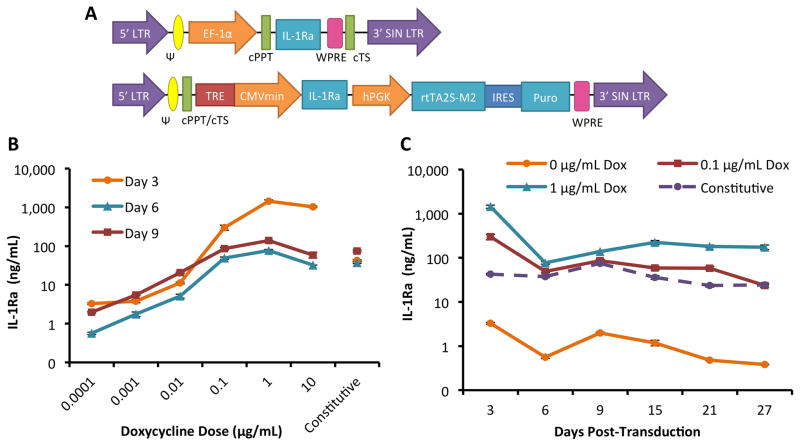
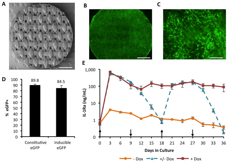
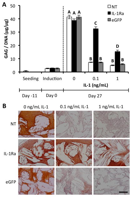
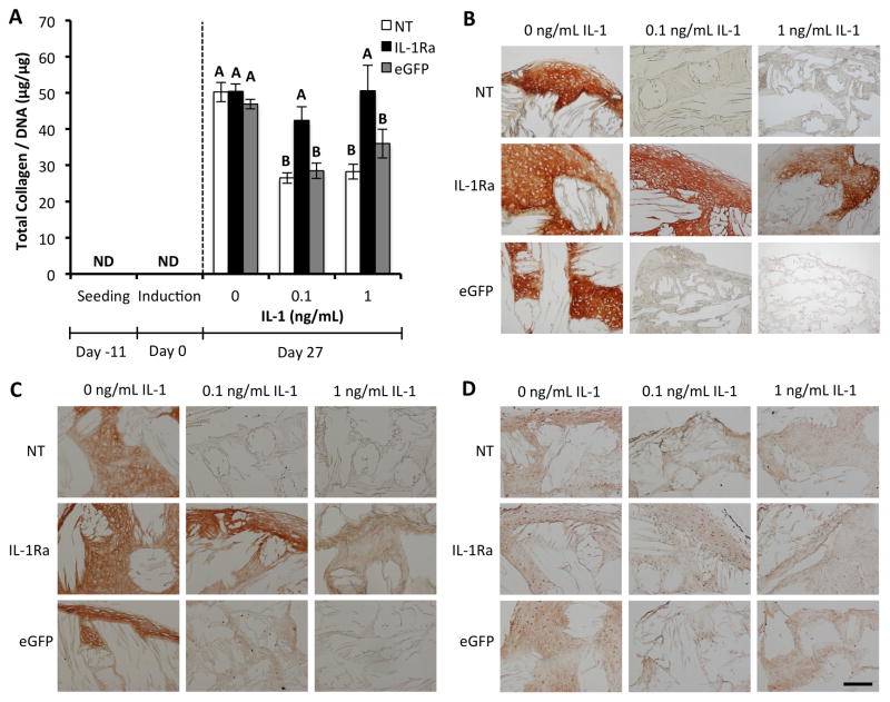
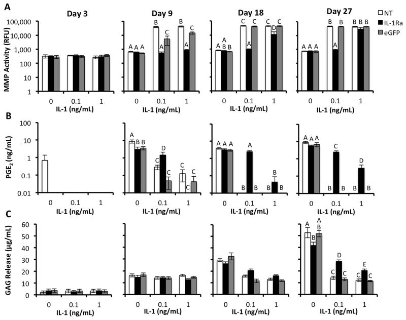
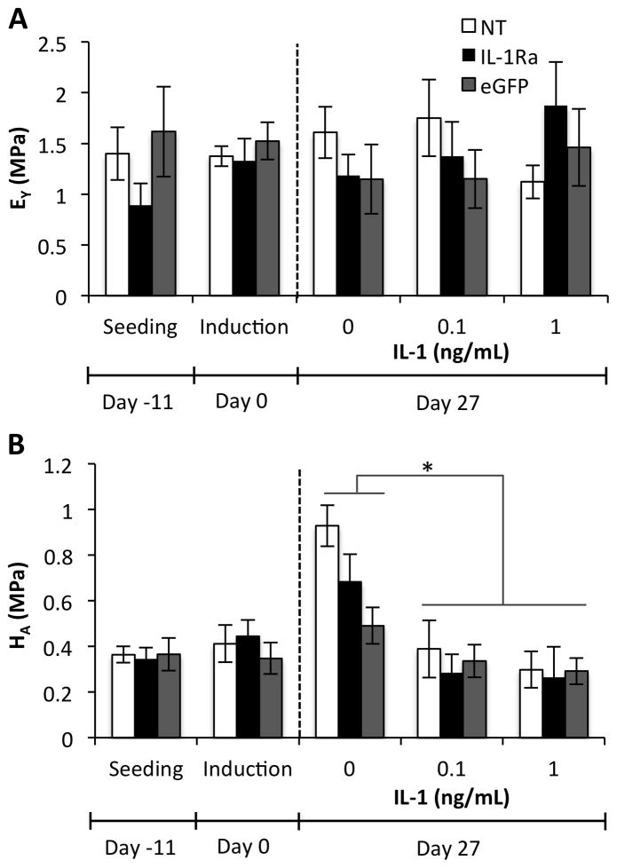
The pathogenesis of osteoarthritis is mediated in part by inflammatory cytokines including interleukin-1 (IL-1), which promote degradation of articular cartilage and prevent human mesenchymal stem cell (MSC) chondrogenesis. In this study, we combined gene therapy and functional tissue engineering to develop engineered cartilage with immunomodulatory properties that allow chondrogenesis in the presence of pathologic levels of IL-1 by inducing overexpression of IL-1 receptor antagonist (IL-1Ra) in MSCs via scaffold-mediated lentiviral gene delivery. A doxycycline-inducible vector was used to transduce MSCs in monolayer or within 3D woven PCL scaffolds to enable tunable IL-1Ra production. In the presence of IL-1, IL-1Ra-expressing engineered cartilage produced cartilage-specific extracellular matrix, while resisting IL-1-induced upregulation of matrix metalloproteinases and maintaining mechanical properties similar to native articular cartilage. The ability of functional engineered cartilage to deliver tunable anti-inflammatory cytokines to the joint may enhance the long-term success of therapies for cartilage injuries or osteoarthritis.

Keywords: Bioactive biomaterial; Cartilage repair; Immunoengineering; Lentivirus; Mesenchymal stem cell; Osteoarthritis.


Figures

Figure 1
Figure 1
Tunable IL-1Ra expression in MSCs with a dox-inducible lentiviral vector. A. Schematic diagram of lentiviral vectors with IL-1Ra as the representative gene of interest. On top is the constitutive expression system driven by the elongation factor 1 alpha (EF-1α) promoter. Shown on bottom is the dox-inducible expression system. IL-1Ra is driven by the tetracycline-regulated minimal CMV promoter (TRE-CMVmin). The human phosphoglycerate kinase (hPGK) promoter constitutively drives expression of the tet-responsive transactivator (rtTA2S-M2) and then, following an internal ribosomal entry site (IRES), the puromycin resistance gene (puro) which enables selection of transduced cells. Both vectors contain the 5′ and 3′ long-terminal repeats (LTR), psi packaging signal (Ψ), the central polypurine tract (cPPT), central termination sequence (cTS), and the woodchuck hepatitis virus post-transcriptional regulatory element (WPRE). B. IL-1Ra secretion tuned by dox dose from MSCs in monolayer culture. Shown is IL-1Ra secreted over 72 hours into culture medium on days 3, 6, and 9 following transduction (mean ± SEM, n=3). C. IL-1Ra secretion from 3 to 27 days in monolayer culture at 3 dox doses or with constitutive expression. Shown is IL-1Ra secreted over 72 hours into culture medium (mean ± SEM, n=3).
Figure 2
Figure 2
Scaffold-mediated LV transduction of MSCs within 3D woven PCL scaffolds. A. Scanning electron microscopy (SEM) image of a 3D woven PCL scaffold 5 mm disk. Scale bar = 1mm. B. Fluorescence image of constitutive eGFP-expressing MSCs on the 3D woven PCL scaffold 6 days after seeding. Scale bar = 1 mm, 300 ms exposure. C. Fluorescence image of constitutive eGFP-expressing MSCs on the 3D woven PCL scaffold 6 days after seeding. Scale bar = 250 μm, 90 ms exposure. D. Scaffold-mediated transduction efficiency of MSCs with either constitutive or inducible eGFP-LV. MSCs were isolated from constructs at day 6 and the percentage of eGFP+ cells was measured via flow cytometry (mean ± SEM, n=3, all samples given 1 μg/mL dox). E. IL-1Ra secretion from engineered cartilage constructs into media every 72 hours over 36 days of chondrogenesis (mean ± SEM, n=3). + Dox indicates dox induction at 1 μg/mL for 36 days. − Dox indicates the baseline IL-1Ra expression in the absence of dox. +/− Dox indicates that dox (1 μg/mL) was switched on and off every 9 days. Upward arrows show time points at which dox was induced and downward arrows show the withdrawal of dox.
Figure 3
Figure 3
IL-1Ra-expressing constructs maintain GAG content with IL-1 treatment. A. GAG per DNA content in engineered cartilage constructs at seeding, chondrogenic induction, and after 27 days of culture in chondrogenic media with either 0, 0.1, or 1 ng/mL IL-1 (mean ± SEM, n=5). NT indicates non-transduced constructs. IL-1Ra indicates constructs transduced with IL-1Ra LV. eGFP indicates constructs transduced with eGFP LV. Groups with different letters are significantly different (P<0.05) by ANOVA and Fisher’s LSD post-hoc. B. Safranin-O red and fast green staining for GAGs and collagen, respectively. The white space shows the location of the PCL fiber bundles. All sections 8 μm. Scale bar = 200 μm.
Figure 4
Figure 4
IL-1Ra-expressing constructs maintain collagen content with IL-1 treatment. A. Collagen per DNA content in engineered cartilage constructs at seeding, chondrogenic induction, and after 27 days of culture in chondrogenic media with either 0, 0.1, or 1 ng/mL IL-1 (mean ± SEM, n=5). NT indicates non-transduced constructs. IL-1Ra indicates constructs transduced with IL-1Ra LV. eGFP indicates constructs transduced with eGFP LV. Groups with different letters are significantly different (P<0.05) by ANOVA and Fisher’s LSD post-hoc. ND = nondetectable. Immunohistochemistry staining for B. Type II collagen. C. Type I collagen. D. Type X collagen. All sections 8 μm. Scale bar = 200 μm.
Figure 5
Figure 5
Analyses of inflammatory mediators and ECM released into culture media by engineered cartilage constructs during chondrogenesis (days 3,9,18,27) and treatment with either 0, 0.1, or 1 ng/mL IL-1. NT indicates non-transduced constructs. IL-1Ra indicates constructs transduced with IL-1Ra LV. eGFP indicates constructs transduced with eGFP LV. For all analyses, groups with different letters are significantly different (P<0.05) by ANOVA and Fisher’s LSD post-hoc (mean +/− SEM, n=5). A. Total specific activity of MMPs released into culture media. B. Concentration of PGE2 released into culture media. c. Concentration of GAG released into culture media.
Figure 6
Figure 6
Mechanical properties of engineered cartilage constructs at seeding, chondrogenic induction, and after 27 days of culture in chondrogenic media with either 0, 0.1, or 1 ng/mL IL-1 (mean ± SEM, n=5). NT indicates non-transduced constructs. IL-1Ra indicates constructs transduced with IL-1Ra LV. eGFP indicates constructs transduced with eGFP LV. A. Equilibrium Young’s modulus (EY). No significant interaction or main effects were found by ANOVA. B. Aggregate modulus (HA). IL-1 treatment was significantly different than no IL-1 treatment at Day 27 (p<0.05), but no significant interaction or main effect of vector was observed by ANOVA.

References

    1. Lawrence RC, Felson DT, Helmick CG, Arnold LM, Choi H, Deyo RA, et al. Estimates of the prevalence of arthritis and other rheumatic conditions in the United States. Part II. Arthritis Rheum. 2008;58(1):26–35. - PMC - PubMed
    1. Abramson SB, Attur M. Developments in the scientific understanding of osteoarthritis. Arthritis Res Ther. 2009;11(3):227. - PMC - PubMed
    1. Anderson DD, Chubinskaya S, Guilak F, Martin JA, Oegema TR, Olson SA, et al. Post-traumatic osteoarthritis: improved understanding and opportunities for early intervention. J Orthop Res. 2011;29(6):802–9. - PMC - PubMed
    1. Chung C, Burdick JA. Engineering cartilage tissue. Adv Drug Deliv Rev. 2008;60(2):243–62. - PMC - PubMed
    1. Johnstone B, Alini M, Cucchiarini M, Dodge GR, Eglin D, Guilak F, et al. Tissue engineering for articular cartilage repair--the state of the art. Eur Cell Mater. 2013;25:248–67. - PubMed

Publication types

Substances