

Papillary renal cell carcinoma revisited: a comprehensive histomorphologic study with outcome correlations
- PMID: 24767860
- PMCID: PMC6180318
- DOI: 10.1016/j.humpath.2014.02.004
Papillary renal cell carcinoma revisited: a comprehensive histomorphologic study with outcome correlations
Abstract
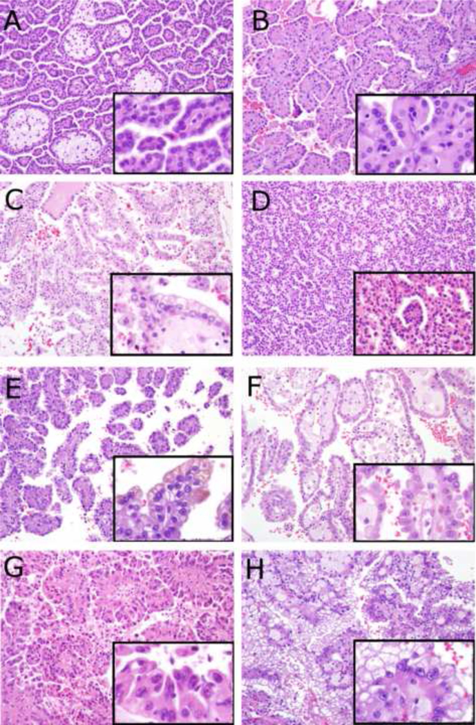
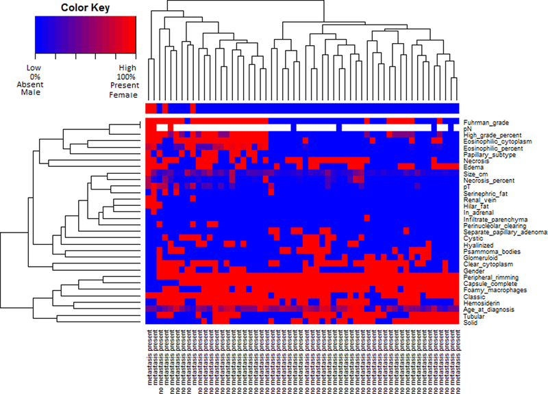
Papillary renal cell carcinoma (P-RCC) is the second most common type of malignant renal epithelial tumor and can be subclassified into type 1, which demonstrates simple cuboidal low-grade epithelium and type 2, which demonstrates pseudostratified high-grade epithelium with abundant eosinophilic cytoplasm. Despite this clinically useful subclassification, P-RCCs exhibit considerable histomorphologic diversity, with many cases having features differing from classically described type 1 and type 2 tumors. To our knowledge, there has been no recent study that has methodically evaluated the histomorphologic features of a series of P-RCCs. To address this, we evaluated a cohort of P-RCCs diagnosed between 1997 and 2004 with long-term clinical follow-up data (n = 56). Histomorphologic features previously described in the spectrum of type 1 and type 2 P-RCCs were recorded for each tumor, including nuclear grade, complete tumor capsule, and cytoplasmic eosinophilia as well as several other features. The current TNM staging (American Joint Committee on Cancer, seventh edition) was assigned to all cases. Histomorphologic features were diverse, demonstrating classic type 1 P-RCC and classic type 2 P-RCC morphology and several tumors with nonclassic features. Four patients in this cohort had distant metastasis. The primary tumor was equally divided between type 1 (2 cases) and type 2 (2 cases) morphology in the cases with metastasis. All P-RCC cases with metastases demonstrated presence of high nuclear grade and high tumor stage in the primary tumor. Cluster analysis using staging parameters and histomorphologic features divided tumors into 2 primary clusters. All primary tumors associated with metastasis were in the same cluster.
Keywords: Nuclear grade; Papillary renal cell carcinoma; Tumor stage.
Copyright © 2014 Elsevier Inc. All rights reserved.
Figures
References
-
- Cheville JC, Lohse CM, Zincke H, Weaver AL, Blute ML. Comparisons of outcome and prognostic features among histologic subtypes of renal cell carcinoma. Am J Surg Pathol. 2003;27:612–24. - PubMed
-
- Delahunt B, Eble JN. Papillary renal cell carcinoma: a clinicopathologic and immunohistochemical study of 105 tumors. Mod Pathol Off J U S Can Acad Pathol Inc. 1997;10:537–44. - PubMed
-
- Eble JN, Sauter G, Epstein JI, Sesterhenn IA. World Health Organization Classification of Tumours of the Urinary System and Male Genital Organs. IARC Press; 2004.
-
- Delahunt B, Eble JN, McCredie MR, Bethwaite PB, Stewart JH, Bilous AM. Morphologic typing of papillary renal cell carcinoma: comparison of growth kinetics and patient survival in 66 cases. Hum Pathol. 2001;32:590–5. - PubMed
-
- Pignot G, Elie C, Conquy S, et al. Survival analysis of 130 patients with papillary renal cell carcinoma: prognostic utility of type 1 and type 2 subclassification. Urology. 2007;69:230–5. - PubMed
Publication types
MeSH terms
Grants and funding
LinkOut - more resources
Full Text Sources
Other Literature Sources
Medical
