. 2014 May 19;24(10):1101-6.
doi: 10.1016/j.cub.2014.03.040. Epub 2014 Apr 24.

Replicative stress induces intragenic transcription of the ASE1 gene that negatively regulates Ase1 activity

Affiliations

Replicative stress induces intragenic transcription of the ASE1 gene that negatively regulates Ase1 activity

Kelly McKnight et al. Curr Biol. .

Abstract

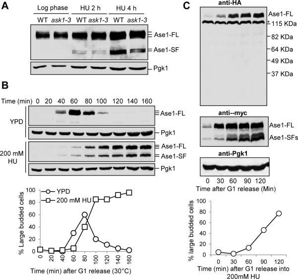
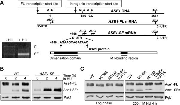
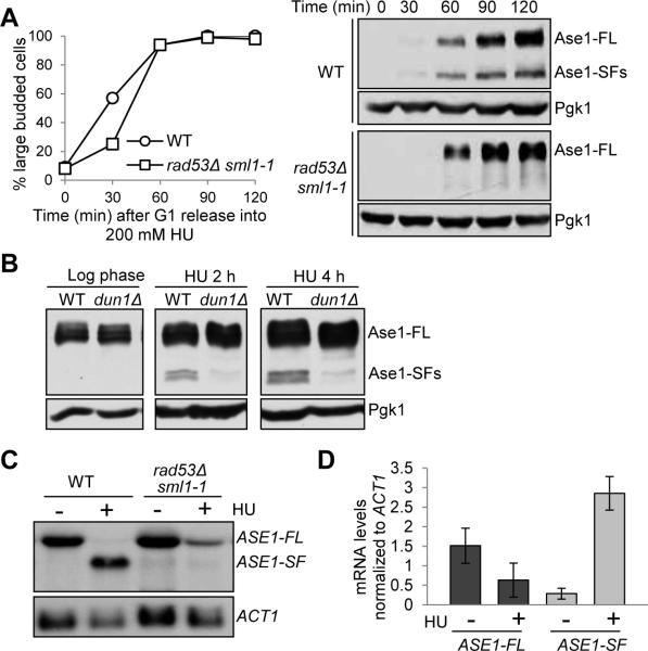
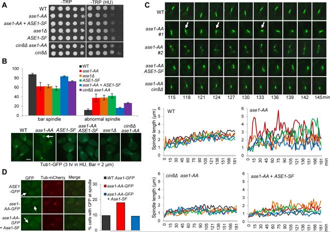
Intragenic transcripts initiate within the coding region of a gene, thereby producing shorter mRNAs and proteins. Although intragenic transcripts are widely expressed [1], their role in the functional regulation of genes remains largely unknown. In budding yeast, DNA replication stress activates the S phase checkpoint that stabilizes replication forks and arrests cells in S phase with a short spindle [2-4]. When yeast cells were treated with hydroxyurea (HU) to block DNA synthesis and induce replication stress, we found that Ase1, a conserved spindle midzone protein [5], appeared as two short protein isoforms in addition to the full-length protein. We further demonstrated that the short isoforms result from intragenic transcription of ASE1, which depends on the S phase checkpoint. Blocking generation of the short isoforms leads to a destabilized S phase spindle, characterized by increased spindle dynamics and frequent spindle collapse. Because the short Ase1 isoforms localize at the spindle in HU-treated cells and overexpression of the short Ase1 isoforms impairs the spindle midzone localization of full-length Ase1, it is likely that the presence of short Ase1 isoforms stabilizes the spindle by antagonizing full-length Ase1. Together, our results reveal intragenic transcription as a unique mechanism to downregulate gene functions in response to DNA replication stress.


Figures

Figure 1
Figure 1. HU-induced expression of Ase1 short protein fragments
(A) The expression of Ase1 in HU-treated WT and ask1-3 mutant cells. ASE1-13myc and ask1-3 ASE1-13myc cells were grown to log phase at 30°C and released to YPD containing 200 mM HU. Cells were collected at the indicated time points and protein samples were prepared for western blotting. Full length Ase1 migrates around 140 kDa and the short isoforms migrate around 105 kDa. Pgk1 protein migrates at 45 kDa and levels are shown as a loading control. (B) Expression of Ase1 isoforms in synchronized cells treated with HU. G1-arrested ASE1-13myc cells were released into YPD or YPD containing 200 mM HU. α-factor was restored in untreated cells to block the second round of cell cycle. Cells were collected at the indicated time points to detect the expression of Ase1 protein using western blotting. Budding index was used to indicate cell cycle stage and Pgk1 protein levels are shown as a loading control. (C) Ase1 short isoforms are not protein cleavage products. G1-arrested ase1Δ cells containing 3HA-ASE1-13myc plasmid were released into fresh selective media containing 200 mM HU and protein samples were prepared at the indicated time points. Western blotting with anti-HA and anti-myc antibodies was used to determine the expression of full length (Ase1-FL) and short isoforms (Ase1-SF) of Ase1 protein. Budding index was used to indicate cell cycle stage and Pgk1 protein levels are shown as a loading control. Asterisk indicates a non-specific band. Sell also Figure S1.
Figure 2
Figure 2. The short Ase1 protein isoforms are a consequence of intragenic transcription
(A) Transcription of ASE1 short mRNA isoform begins at bp +756 to +766. Gel image for ASE1 gene specific PCR products after 5-RACE. Schematic of results from 5-RACE after cloning and sequencing of PCR bands (10 colonies each). (B) Full-length ASE1 mRNA is not required for expression of Ase1-SFs. ase1Δ cells containing WT ASE1 or ASE1-SF plasmid were grown to log phase at 30°C and released into selective media containing 200 mM HU. Cells were collected at the indicated time points and protein samples were prepared for western blotting to determine the expression of Ase1-FL and Ase1-SFs. (C) Translation of Ase1-SFs starts at M286 and M313. ase1Δ cells containing ASE1-13myc, ase1M286A-13myc, ase1M313A-13myc or ase1M286A-M313A-13myc plasmid were grown to log phase in selective media and released into 200 mM HU for 4 hrs. Cells were collected and protein samples were prepared for western blotting with anti-myc antibody. Pgk1 protein levels are shown as a loading control. See also Figure S2.
Figure 3
Figure 3. The expression of the Ase1 short isoforms depends on the S-phase checkpoint
(A) Rad53 is required for Ase1 short protein isoform expression. G1-arrested ASE1-13myc and rad53Δ sml1-1 ASE1-13myc cells were released into YPD containing 200 mM HU. Cells were collected at the indicated time points and protein samples were prepared for western blotting. Budding index was used to indicate cell cycle stage. (B) The expression of Ase1 isoforms in dun1Δ mutants. ASE1-13myc and dun1Δ ASE1-13myc cells were grown to log phase at 30°C and released into YPD containing 200 mM HU. Cells were collected at the indicated time points and protein samples were prepared for western blotting to determine the expression of Ase1 protein. Pgk1 protein levels are shown as a loading control. (C) Intragenic transcription of ASE1 depends on Rad53. G1-arrested WT and rad53Δ sml1-1 cells were released to fresh YPD at 30°C. Twenty min later 200 mM HU was added to half of the cell culture. Cells were harvested 40 min later, washed with 1xPBS and flash frozen with liquid nitrogen. mRNA was prepared and examined by northern blotting with a probe corresponding to nucleotides 2109 to 2411 of the ASE1 gene. ACT1 probe was used for loading control. (D) Quantification of the ASE1 mRNAs in WT cells treated with or without HU from three experiments normalized to ACT1 mRNA using ImageJ software: ASE1-FL –HU (1.51 ± 0.45), ASE1-FL + HU (0.63 ± 0.43), ASE1-SF – HU (0.26 ± 0.14), ASE1-SF + HU (2.86 ± 0.43). See also Figure S3.
Figure 4
Figure 4. Expression of the Ase1 short isoforms stabilizes the S-phase spindle
(A) ase1-AA mutants are sensitive to HU. Saturated cell cultures with indicated genotypes were 10-fold serial diluted and spotted onto –TRP and –TRP + 100 mM HU plates and scanned after incubation at 30°C for 2 days. (B) ase1-AA cells show abnormal spindles after HU treatment. Cells with indicated genotypes were arrested in G1 and released to fresh selective media containing 200 mM HU for 3 hrs at 30°C. The spindle morphology (Tub1-GFP) in representative cells is shown in the bottom panel. The white arrow indicates a collapsed spindle. The percentage of cells with a normal bar-shaped spindle and an abnormal collapsed or dot-like spindle from three biological replicates is shown in the top panel (n > 100). Normal: WT 88% ± 2.3, ase1-AA 62.3% ± 8.2, ase1Δ 62.6% ± 3.6, ASE1-SF 57.9% ± 4.4, ase1-AA + ASE1-SF 83.7% ± 1.3, cin8Δ ase1-AA 73.2% ± 1.5. Abnormal: WT 12% ± 2.3, ase1-AA 37.7% ± 8.2, ase1Δ 38.4% ± 5.1, ASE1-SF 42.1% ± 4.4, ase1-AA + ASE1-SF 16.3% ± 1.3, cin8Δ ase1-A 26.8% ± 1.5. (C) Spindles are more dynamic and experience more collapse in ase1-AA mutants during HU treatment. Thirty min following G1 release, the cells with indicated genotypes were spotted onto the surface of a slide with an agarose medium pad containing 200 mM HU and subjected to live-cell microscopy. Five representative cells for each genotype were selected and spindle length was measured using Andor iQ2 software. Time zero was set to one image prior to the first separation of spindle poles and spindle length was plotted over time (bottom panel). Live-cell images of the spindle morphology in representative cells are shown in the top panel. Arrows indicate spindle collapse. See also Figure S4. (D) Ase1-SF reduces Ase1-FL spindle localization. G1-arrested cells with indicated genotypes were released to liquid media containing 200 mM HU for 1 hr, and then mounted to a slide as described in (C). The cells were subjected to live-cell microscopy for 30 min after G1 release for 2 hrs. The percentage of cells showing GFP localization to the spindle in several time points during the 30 min window is shown (n >100). See also Figure S4.

Similar articles

Cited by

References

    1. Pelechano V, Wei W, Steinmetz LM. Extensive transcriptional heterogeneity revealed by isoform profiling. Nature. 2013;497:127–131. - PMC - PubMed
    1. Bachant J, Jessen SR, Kavanaugh SE, Fielding CS. The yeast S phase checkpoint enables replicating chromosomes to bi-orient and restrain spindle extension during S phase distress. J Cell Biol. 2005;168:999–1012. - PMC - PubMed
    1. Alcasabas AA, Osborn AJ, Bachant J, Hu F, Werler PJ, Bousset K, Furuya K, Diffley JF, Carr AM, Elledge SJ. Mrc1 transduces signals of DNA replication stress to activate Rad53. Nat Cell Biol. 2001;3:958–965. - PubMed
    1. Li Y, Bachant J, Alcasabas AA, Wang Y, Qin J, Elledge SJ. The mitotic spindle is required for loading of the DASH complex onto the kinetochore. Genes Dev. 2002;16:183–197. - PMC - PubMed
    1. Schuyler SC, Liu JY, Pellman D. The molecular function of Ase1p: evidence for a MAP-dependent midzone-specific spindle matrix. Microtubule-associated proteins. J Cell Biol. 2003;160:517–528. - PMC - PubMed

Publication types

MeSH terms

LinkOut - more resources