. 2014 May 6;19(5):836-48.
doi: 10.1016/j.cmet.2014.03.023. Epub 2014 Apr 24.

PTENα, a PTEN isoform translated through alternative initiation, regulates mitochondrial function and energy metabolism

Affiliations

PTENα, a PTEN isoform translated through alternative initiation, regulates mitochondrial function and energy metabolism

Hui Liang et al. Cell Metab. .

Abstract

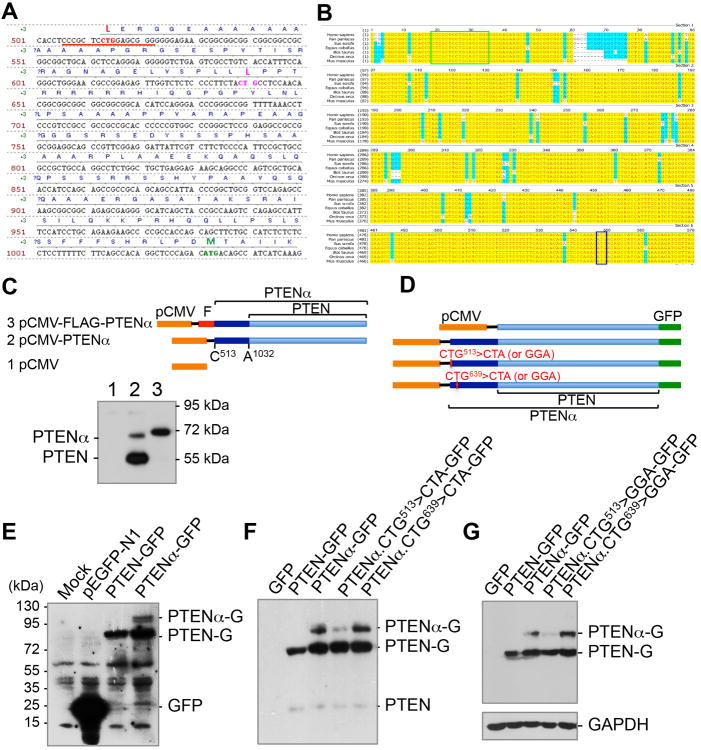
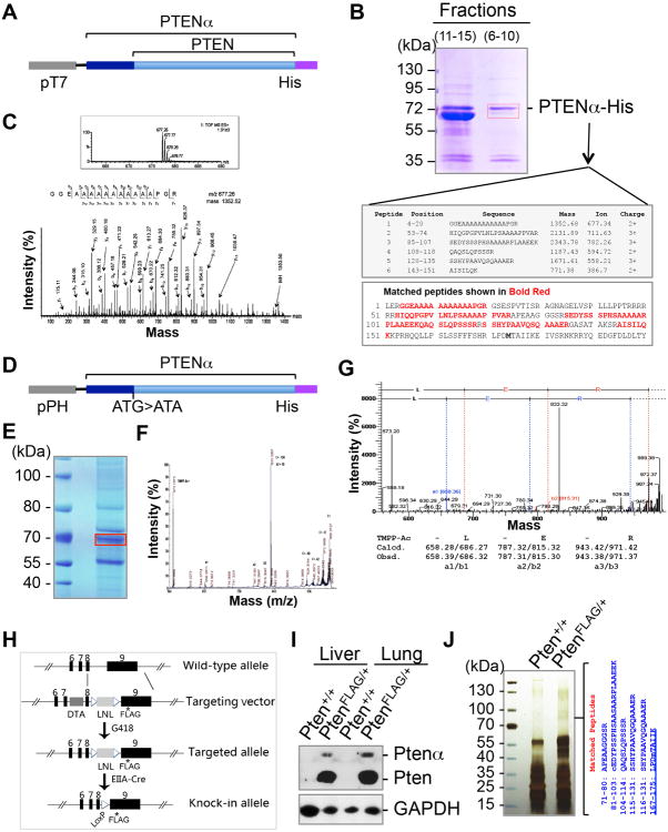
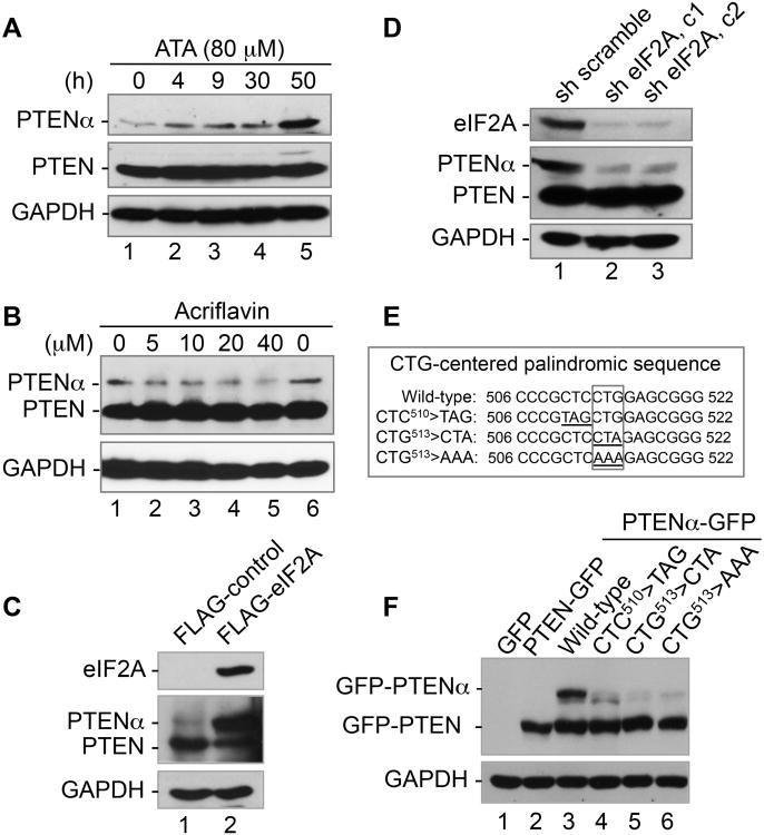
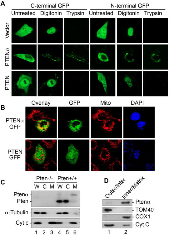
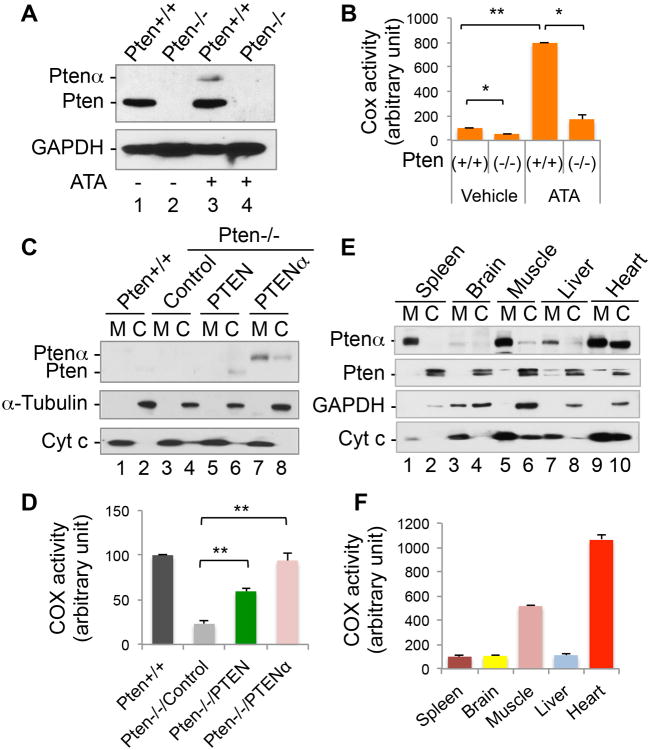
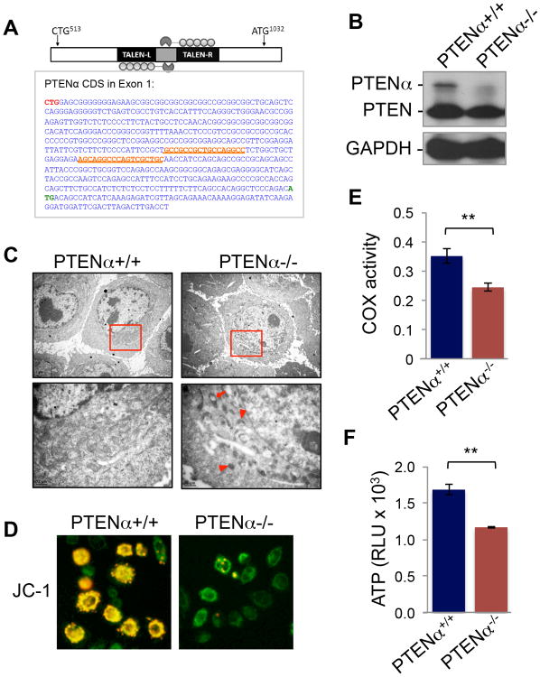
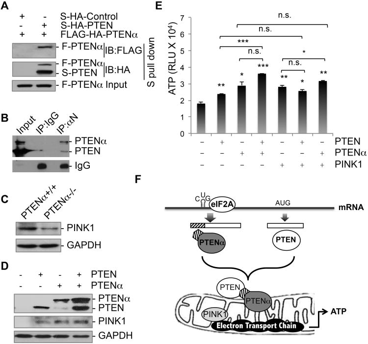
PTEN is one of the most frequently mutated genes in human cancer. It is known that PTEN has a wide range of biological functions beyond tumor suppression. Here, we report that PTENα, an N-terminally extended form of PTEN, functions in mitochondrial metabolism. Translation of PTENα is initiated from a CUG codon upstream of and in-frame with the coding region of canonical PTEN. Eukaryotic translation initiation factor 2A (eIF2A) controls PTENα translation, which requires a CUG-centered palindromic motif. We show that PTENα induces cytochrome c oxidase activity and ATP production in mitochondria. TALEN-mediated somatic deletion of PTENα impairs mitochondrial respiratory chain function. PTENα interacts with canonical PTEN to increase PINK1 protein levels and promote energy production. Our studies demonstrate the importance of eIF2A-mediated alternative translation for generation of protein diversity in eukaryotic systems and provide insights into the mechanism by which the PTEN family is involved in multiple cellular processes.


Figures

Figure 1
Figure 1. Identification and validation of PTENα
(A) Sequence of the 5′-UTR region of Homo sapiens PTEN mRNA. Two potential CUG sites (red and pink) as well as the normal ATG start site (green) are highlighted. (B) The 5′-UTR of PTEN containing the 16-bp CUG-centered palindromic motif is evolutionary conserved. Phylogenetic analysis of the 5′-UTR of PTEN mRNA in bonobo (Pan paniscus), wild boar (Sus scrofa), horse (Equus caballus), cattle (Bos taurus), killer whale (Orcinus orca) and mouse (Mus musculus). The 16-bp palindromic sequence is highlighted in a green box and the ATG start codon of canonical PTEN is in a blue box. (C) PTEN cds with a CUG513-starting 5′-UTR region (PTENα) was constructed under a CMV promoter and expressed with and without a N-terminal FLAG tag. Human HEK293T cells were transfected with indicated expression plasmids, followed by Western blotting analysis using a PTEN monoclonal antibody against its C-terminal region. (D) A different set of constructs of PTEN and PTENα with a C-terminal GFP tag, in which one of the two possible CTG sites (CTG513 or CTG639) was mutated to CTA or GGA. (E-G) Mutation of CTG513 but not CTG639 eliminates the expression of PTENα. C-terminal GFP-tagged PTENα expression plasmids with and without CTG513>CTA or CTG513>GGA mutation(s) were introduced in human HCT116 colon cancer cells, followed by detection of GFP-PTENα variants. E, The expression of GFP-tagged PTEN or PTENα was confirmed by Western blotting using a GFP antibody. F and G, mutation of CTG513 but not CTG639 into CTA or GGA results in the disappearance of PTENα. PTENα-G and PTEN-G, PTENα or PTEN with a C-terminal GFP tag. See also Figure S1.
Figure 2
Figure 2. The translation initiation codon of PTENα is identified by MALDI-TOF mass spectrometry and terminal analysis with de novo sequencing
(A) A pET28a plasmid containing PTENα with a C-terminal His-tag used for in vitro purification and mass spectrometry sequencing. (B) His-selected affinity purification of PTENα. Bacteria-expressed His-PTENα was purified using nickel affinity chromatography. Combined fractions containing the slow migrating PTEN band (fractions 6-10) confirmed by PTEN immunoblotting (not shown) were separated with SDS-PAGE. Mass spectrometry analysis of a purified ∼70 kDa band (in red box) revealed six pieces of peptide that match the 5′-UTR region of PTEN, including a peptide near the CTG513-leucine site. (C) The MS/MS spectrum of the peptide (GGEAAAAAAAAAAAPGR) that matches the N-terminus sequence of PTENα. (D) A pFastBac1 plasmid containing PTENα with a C-terminal His-tag used for in vitro purification and mass spectrometry sequencing. The PTEN ATG start codon was mutated to ATA to avoid PTEN co-purification with PTENα. (E) Purification of SF9-expressed PTENα with a C-terminal His-tag (band in red box) forMALDI-TOF-TOF MS and TMPP-Ac-OSu derivatization for de novo sequencing. (F) Tandem spectrum of m/z 989.3727 in TMPP-Ac derivatized PTENα. (G) De novo analysis of m/z 989.35 showing the first amino acids of PTENα, Leucine-Glutamic acid-Arginine (LER). (H) Generation of Pten C-terminal FLAG knock-in mice for verification of the Ptenα gene locus. (I) Verification of expression of FLAG-tagged PTEN and PTENα in Pten-FLAG knock-in mice. Liver and lung tissues from Pten-FLAG knock-in mice or control wild-type mice were lysed for immunoblotting with anti-FLAG antibody. (J) Protein lysates of Pten-FLAG knock-in liver tissues or control tissues were subjected to sequential immunoprecipitation with anti-FLAG M2 agarose and a PTENα-specific antibody. The bound proteins were separated with SDS-PAGE and gel slices at around 70-kDa were analyzed by mass spectrometry. Four peptides were identified in Pten-FLAG knock-in tissues that match the N-terminally extended region of PTENα, αN. See also Figure S2.
Figure 3
Figure 3. PTENα is synthesized through an eIF2A-mediated CUG initiation mechanism and a palindrome sequence is essential for PTENα expression
(A) Induction of PTENα by aurin tricarboxylic acid (ATA) in a time-dependent manner. HeLa cells were treated with ATA (80 mM) for various periods of time and the expression of PTENα as well as PTEN was examined by Western blot. (B) Dose-dependent inhibition of PTENα expression by acriflavin. HeLa cells were treated with different doses of acriflavin for 4 h prior to immunoblotting for evaluation of PTENα expression. Expression levels of PTEN and GAPDH were included as controls. (C) eIF2A alters the ratio of PTENα versus PTEN by up-regulating PTENα and down-regulating PTEN. FLAG-tagged eIF2A was overexpressed in HEK293T cells prior to Western evaluation of PTENα and PTEN. eIF2A expression was verified by probing the same blot with anti-FLAG antibody. GAPDH was used as a loading control. (D) Reduction of PTENα in response to knockdown of eIF2A. HeLa cells were infected with lentivirus expressing shRNA of eIF2A or scramble shRNA. Cell lysates were analyzed by Western blotting with antibodies against eIF2A, PTEN (m) and GAPDH. (E) CTG513-centered palindromic sequence and mutagenesis disruption of the palindrome. (F) Abolition of PTENα by palindrome disruption. Mutations were made at CTC510, thetriplet immediately before the CTG513 start codon, or at CUG513 itself as indicated prior to Western analysis of PTENα expression. See also Figure S3.
Figure 4
Figure 4. PTENα is localized predominantly in cytoplasm and mitochondria
(A) Pten-/- MEFs transfected with N-terminal or C-terminal GFP-tagged PTEN or PTENα were subjected to protease protection assay, confirming the difference in subcellular distribution patters of PTENα and PTEN. (B) Subcellular localization of C-terminal GFP-tagged PTENα (with an ATG>ATA mutation) and PTEN shown by confocal fluorescence microscopy. MitoTracker was used to indicate mitochondria. Overlay, merged images of GFP and MitoTracker. (C) Cell fractionation was performed to isolate mitochondria in Pten+/+ and Pten-/- MEFs prior to immunoblotting analysis of PTENα and PTEN expression. W, whole cell lysate; M, mitochondria; C, cytoplasm. Cytochrome c and a-tubulin were used as mitochondrial and cytoplasmic markers. (D) Mitochondria isolated from mouse brain cortex were subjected to sub fractionation of mitochondria prior to evaluation of PTENα by immunoblotting. Tom40 and Cox1 were used as markers for the mitochondrial outer membrane and inner membrane respectively. Cytochrome C is a dynamic component of mitochondria and can be found in both the inner membrane and intermembrane space. See also Figures S4-S7.
Figure 5
Figure 5. PTENα regulates cytochrome c oxidase activity
(A) Pten+/+ and Pten -/- MEFs were treated with ATA (100mM, 24 h) and examined for expression levels of PTEN and PTENα. (B) Mitochondrial fractions were extracted from Pten+/+ and Pten-/- MEFs treated with ATA as in (A) for analysis of cytochrome c oxidatase (COX) activity. Data are presented as mean±SEM of three independent experiments and analyzed with the paired t-test. *, p <0.05; **, p <0.01. (C) Pten+/+ and Pten-/- MEFs with and without ectopic expression of FLAG-tagged PTEN or PTENα were subjected to a cell fractionation procedure for isolation of mitochondria, followed by immunoblot analysis of PTEN/PTENα expression. Cytochrome c and α-tubulin were used as mitochondrial and cytoplasmic markers. M, mitochondria; C, cytoplasm. (D) Mitochondria from Pten+/+ MEFs as well as from Pten-/- MEFs containing ectopic PTEN or PTENα were analyzed for COX activity. Data are presented as mean±SEM of three independent experiments and analyzed by paired t-test. **, p <0.01. (E) Various mouse tissues were subjected to mitochondria/cytoplasm fractionation, followed by Western analysis of PTENα expression. PTEN expression is also shown for comparison. GAPDH and cytochrome c were used as cytoplasmic and mitochondrial markers. (F) Mitochondria were extracted from various mouse tissues as indicated for analysis of COX activity. See also Figures S4-S7.
Figure 6
Figure 6. Somatic knockout of PTENα impairs mitochondrial structure and function
(A) Somatic knockout of PTENα with the TALEN technique. Upper panel, schematic strategy of PTENα TALEN knockout. Lower panel, sequence of PTENα CDS in exon 1. TALEN targeted left and right arms are underlined and highlighted in orange. CTG513 and ATG1032 are highlighted in red and green respectively. (B) Western blot confirming elimination of PTENα. (C) Marked mitochondrial morphological alterations in PTENα-/- HeLa cells shown by electron microscopy. Arrowheads indicate smaller condensed mitochondria. The arrow points to a mitochondrion with expanded vesicles. (D) JC-1 staining showing loss of red-J-aggregate fluorescence in PTENα-/- cells (right) as compared with PTENα+/+ cells (left). (E) Impaired COX activity in PTENα-/- cells. Mitochondria were extracted from PTENα+/+ and PTENα-/- cells for analysis of COX activity. (F) PTENα knockout reduces ATP production. Data are presented as mean±SD of three replicates and analyzed by paired t-test. **, p <0.01.
Figure 7
Figure 7. PTENα and PTEN form a complex and collaborate in energy metabolism
(A) S-HA-tagged PTEN and FLAG-HA-tagged PTENα were transfected into 293T cells prior to S protein pull-down (S-PD). FLAG or HA immunoblotting was performed to detect PTEN-associated PTENα. (B) In vivo binding of PTENα with PTEN. Endogenous PTENα was immunoprecipited using anti-αN antibody from mouse brain tissues for immunoblotting of PTEN. (C) Evaluation of PINK1 expression in PTENα depleted cells by Western blotting. (D) PINK1 expression was assessed in Pten-/- MEFs transfected with PTEN, PTENα, or PTEN+PTENα. (E) Pten-/- MEFs transfected individually or in different combinations with PTEN, PTENα and PINK1, followed by analysis of ATP production. Data are presented as mean±SD. Labeling for statistical significance above each column indicates a comparison with the control column. n.s. not significant, p>0.05; *,p <0.05; **, p <0.01; ***, p <0.001. (F) A graphic model of PTENα translation and its function in mitochrondrial energy metabolism. PTENα is synthesized through an eIF2A- and palindrome-dependent CUG initiation mechanism. PTENα forms a complex with canonical PTEN and these molecules collaborate in mitochondrial bioenergetics through regulation of cytochrome c oxidase activity and ATP production. See also Figures S6 and S7.

Comment in

References

    1. Boch J, Scholze H, Schornack S, Landgraf A, Hahn S, Kay S, Lahaye T, Nickstadt A, Bonas U. Breaking the code of DNA binding specificity of TAL-type III effectors. Science. 2009;326:1509–1512. - PubMed
    1. Chen W, Lee PJ, Shion H, Ellor N, Gebler JC. Improving de novo sequencing of peptides using a charged tag and C-terminal digestion. Anal Chem. 2007;79:1583–1590. - PubMed
    1. Coenen MJ, van den Heuvel LP, Smeitink JA. Mitochondrial oxidative phosphorylation system assembly in man: recent achievements. Curr Opin Neurol. 2001;14:777–781. - PubMed
    1. Di Cristofano A, Pesce B, Cordon-Cardo C, Pandolfi PP. Pten is essential for embryonic development and tumour suppression. Nat Genet. 1998;19:348–355. - PubMed
    1. Fang M, Shen Z, Huang S, Zhao L, Chen S, Mak TW, Wang X. The ER UDPase ENTPD5 promotes protein N-glycosylation, the Warburg effect, and proliferation in the PTEN pathway. Cell. 2010;143:711–724. - PubMed

Publication types