. 2014 Jul:17:132-40.
doi: 10.1016/j.mito.2014.04.008. Epub 2014 Apr 24.

ERK-mediated phosphorylation of TFAM downregulates mitochondrial transcription: implications for Parkinson's disease

Affiliations

ERK-mediated phosphorylation of TFAM downregulates mitochondrial transcription: implications for Parkinson's disease

Kent Z Q Wang et al. Mitochondrion. 2014 Jul.

Abstract

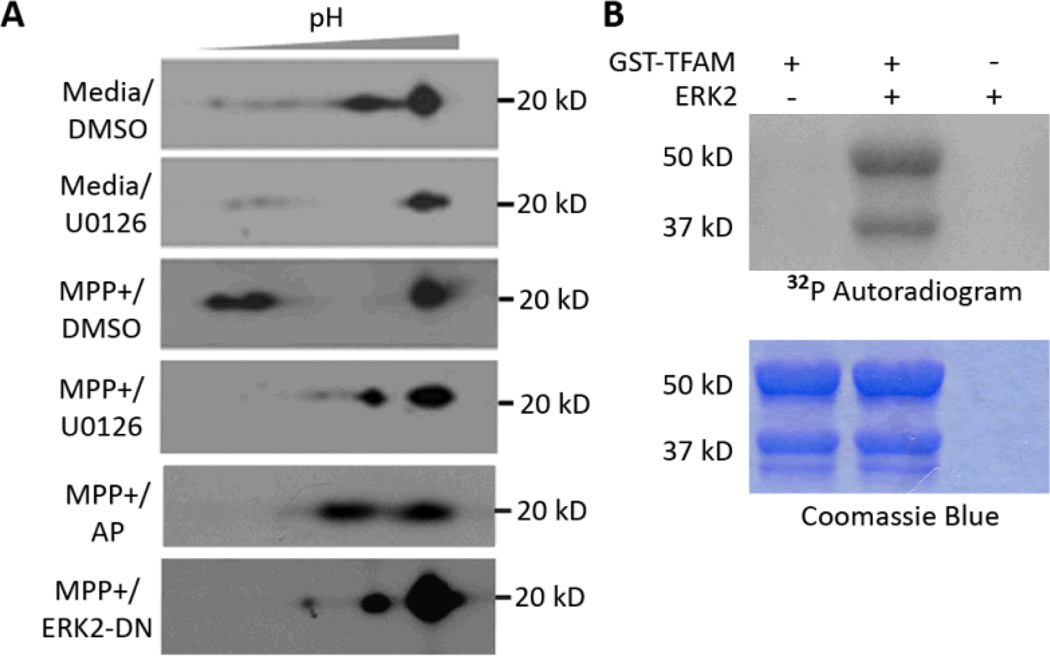
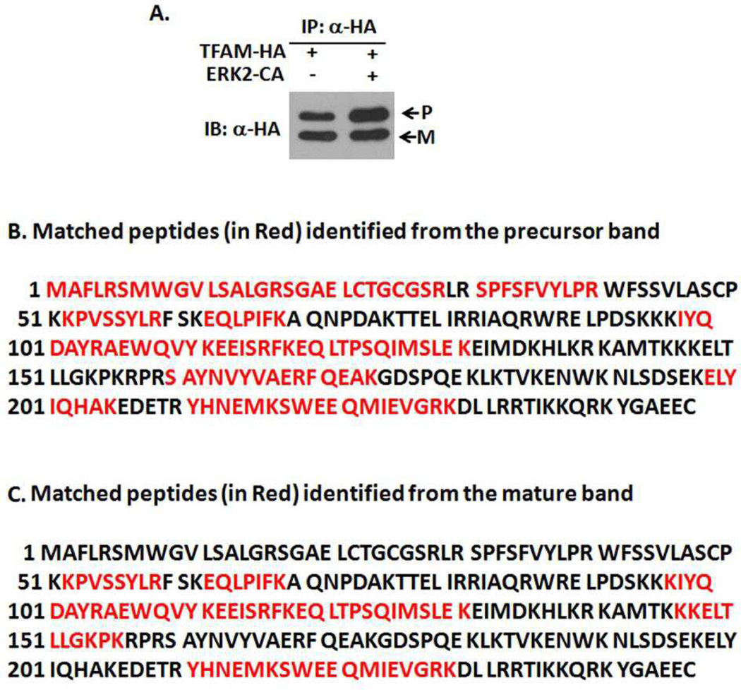
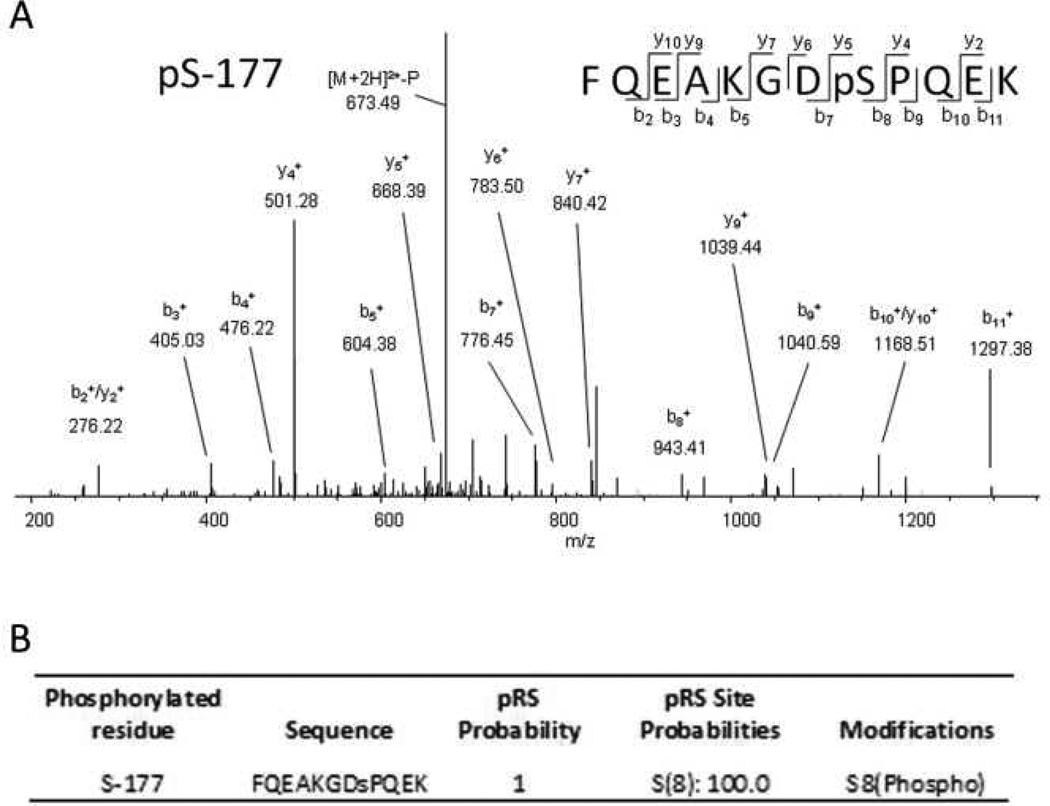
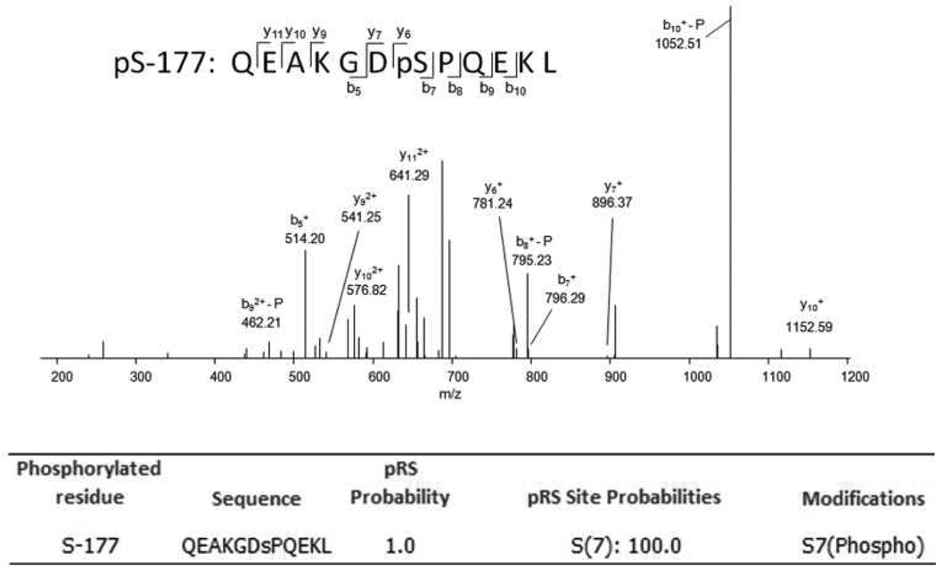
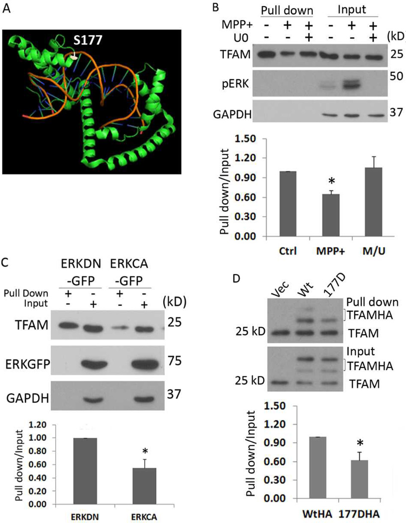
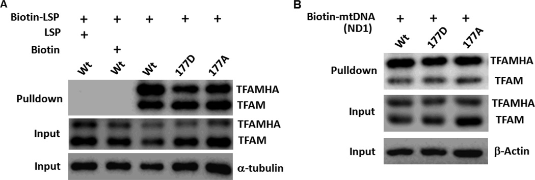
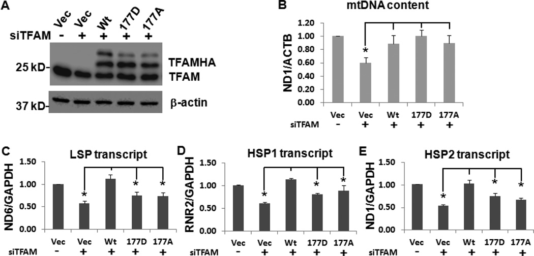
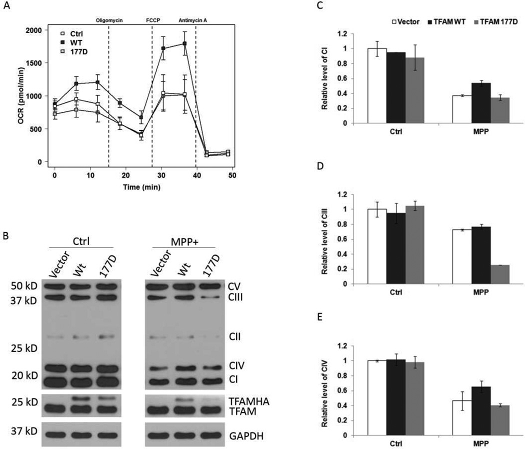
Mitochondrial transcription factor A (TFAM) regulates mitochondrial biogenesis, which is downregulated by extracellular signal-regulated protein kinases (ERK1/2) in cells treated chronically with the complex I inhibitor 1-methyl-4-phenylpyridinium (MPP+). We utilized mass spectrometry to identify ERK1/2-dependent TFAM phosphorylation sites. Mutation of TFAM at serine 177 to mimic phosphorylation recapitulated the effects of MPP+ in decreasing the binding of TFAM to the light strand promoter, suppressing mitochondrial transcription. Mutant TFAM was unable to affect respiratory function or rescue the effects of MPP+ on respiratory complexes. These data disclose a novel mechanism by which ERK1/2 regulates mitochondrial function through direct phosphorylation of TFAM.

Keywords: MAP kinases; Mitochondrial biogenesis; Parkinson disease; Phosphorylation; mtDNA.


Figures

Fig. 1
Fig. 1
ERK-dependent phosphorylation of TFAM. (A) Retinoic acid-differentiated SH-SY5Y cells were treated with the indicated reagents for 6 days prior to analysis. TFAM post-translational modification was analyzed by 2-D immunoblotting using an anti-TFAM antibody. Media is the negative control for MPP+ and DMSO is the negative control for U0126. MPP+/AP indicates MPP+ treated cell lysates that were incubated in vitro with alkaline phosphatase (AP); MPP+/ ERK2-DN refers to MPP+ treatment of cells expressing a dominant negative ERK2. (B) In vitro phosphorylation was performed using recombinant active ERK2 and GST-TFAM as the substrate in the presence of 32P-labeled ATP and detected using autoradiography. Protein loading was visualized by Coomassie Blue staining.
Fig. 2
Fig. 2
Protein ID of TFAM-HA verified by mass spectrometry. The TFAM-HA plasmid was either overexpressed alone or co-expressed with an ERK2-CA plasmid in SH-SY5Y cells for 48 h. Recombinant TFAM was then immunoprecipitated using anti-HA conjugated magnetic beads. Each eluted protein sample was split for loading in parallel lanes on the same 12% SDS-gel, with one lane loaded with 90% and the other loaded with 10% of the total eluate. (A) After electrophoresis, the gel piece containing 10% of the eluted proteins was transferred to a PVDF membrane and immunoblotted for HA. (B & C) The gel piece loaded with 90% of the eluted proteins was stained with Coomassie blue. The precursor and mature TFAM-HA bands (corresponding to the HA-Tag immunoreactive bands in (A)) were excised for trypsin in-gel digestion, and analyzed using MALDI-TOF/TOF. The matched tryptic peptides of TFAM (UniprotKB/Swiss-Prot access number Q00059) are indicated in red characters.
Fig. 3
Fig. 3
Identification of ERK-directed phosphorylation sites of TFAM in cells. TFAM-HA was co-expressed with either a control plasmid or with a constitutively active ERK2 plasmid (ERK2-CA) in SH-SY5Y cells for 48 h, followed by immunoprecipiration of TFAM-HA. Eluted proteins were resolved by 4–15% SDS-PAGE and tryptic peptides generated by in-gel digestion. (A) MS/MS spectra of ERK2-dependent TFAM phosphopeptides modified at S-177 from the TFAMHA and ERK2-CA co-expressed sample. Peptide sequences were identified using the SEQUEST algorithm. (B) The phosphorylation sites were localized using the PhosphoRS algorithm and site probabilities >90% were considered high confidence. Phosphorylated Residue: Position of phosphorylated Serine (S) or Threonine (T) in TFAM (Q00059); Sequence: amino acid sequence of tryptic peptides with the phosphorylated residue displayed in lowercase; pRS probability: probability that the isoform is correct with a maximum value of 1; pRS Site Probabilities: Percent probability for each possible phosphorylation site on the peptide. Modifications: amino acid residue, position, and type of modification on the peptide.
Fig. 4
Fig. 4
Identification of in vitro ERK2-mediated phosphorylation of TFAM. (A) TFAM in vitro phosphorylation reaction was carried out as described in the Materials and Methods. After the reaction, proteins were loaded into a 4–15% SDS polyacryamide gel as: 90% of the reaction to one lane and another 10% of the reaction to a separate lane. After electrophoresis, the gel was sliced into two pieces. The gel piece loaded with 90% of the proteins was stained with Coomassie blue G250. The major protein bands (50 and 38 kD) were excised for trypsinization and MS analysis. Proteins from the other gel piece were transferred onto a PVDF membrane and immunoblotted with anti-TFAM and anti-ERK1/2. (B) Tryptic peptides prepared from the 50 and 38 KD protein bands were analyzed by MALDI-TOF/TOF-MS. All identified peptide fragments are highlighted in red. (C) Chymotryptic peptides from the 50 KD band (full-length GST-TFAM) were analyzed by LC-MS/MS and phosphorylation at (S177) of TFAM was identified.
Fig. 5
Fig. 5
TFAM binding to LSP is reduced by ERK-mediated phosphorylation at S177. (A) The 3D structure of the TFAM-LSP DNA complex (RSCB: 3TMM) rendered with PyMOL predicts that residue S177 (white) is bound to the minor groove of the LSP DNA. (B) RA differentiated SH-SY5Y cells were treated with the indicated reagents for 6 days. Endogenous TFAM protein from each cell lysate was pulled down with a biotinylated DNA probe derived from the mitochondrial D-loop region (biotin-LSP) and analyzed by Western Blot. (C) Endogenous TFAM binding to the LSP was analyzed in HEK 293 cells transfected 48 h prior with plasmids encoding dominant negative ERK2-GFP (ERK2DN-GFP or ERKDN) or constitutively activated ERK2-GFP (ERK2CA-GFP or ERKCA). (D) The indicated TFAM-HA and mutant plasmids were transfected into HEK 293 cells for 48 h. Biotin-LSP + streptavidin-magnetic beads were used to pull-down TFAM for detection using anti-TFAM antibody. Note reduced pull-down of the 177D mutant despite equivalent expression in the input, and equivalent pull-down of endogenous (wild-type) TFAM (bottom bands in the pull-down) serves as an internal control. The relative TFAM-LSP binding shown in (B–D) was calculated as the ratio of the TFAM band from the pull down normalized to input, and presented as Mean ± SEM of three independent experiments. Asterisks indicate p<0.05 (MPP+ versus vehicle control in B; ERKCA versus ERKDN in C; 177DHA versus WtHA in D).
Fig.6
Fig.6
Relative binding of TFAM mutants to LSP and non-specific mtDNA. HEK 293 cells were transfected with TFAM-HA, TFAM(S177D)-HA and TFAM(S177A)-HA mutant plasmids for 48 h and lysed for further analysis. (A) Biotin-LSP and streptavidin-magnetic beads were used to pull-down TFAM for detection using anti-TFAM antibody similar to the procedure shown in Fig. 5D. To validate the LSP binding specificity to TFAM, either 10 times (molar ratio) of non-labeled LSP or free biotin was pre-incubated with an equal amount of lysate containing wildtype TFAM and streptavidin-magnetic beads for 2 h prior to addition of the biotin-LSP probe. (B) A biotinylated mtDNA probe (biotin-mtDNA) derived from a coding region of the Homo sapiens ND1 was used to pull-down TFAM from cell lysates transfected with TFAM-HA, TFAM(S177D)-HA or TFAM(S177A)-HA. There were no differences in the non-specific mtDNA binding of the tested mutants relative to wild-type TFAM-HA. Representative data from 2 independent experiments is shown. Symbol designations in figure panels: Wt as TFAM-HA, 177D as TFAM(S177D)-HA and 177A as TFAM(S177A)-HA.
Fig. 7
Fig. 7
TFAM(177D)-HA is incapable of rescuing mitochondrial transcription incurred by knockdown of endogenous TFAM. HEK293 cells were first transfected with siCtrl or siTFAM for 24 h to knockdown endogenous TFAM, followed by transfection of TFAM-HA plasmids or control vector for another 48 h. (A) The expression levels of the endogenous TFAM and TFAM-HA wild-type and mutant proteins were analyzed by western blot. (B) mtDNA content analyzed by qPCR for ND1 normalized to ACTB. (C-D) The steady-state levels of mitochondrial transcripts of the light strand promoter (LSP), heavy strand promoter 1 (HSP1), and heavy strand promoter 2 (HSP2) were analyzed by qRT-PCR. Data presented in B-E were averaged from five independent experiments and represented as mean ± SEM. Asterisks indicate p<0.05 compared with the Wt. Symbols presented in figure panels: Vec as vector control, Wt as TFAM-HA, 177D as TFAM(S177D)-HA and 177A as TFAM(S177A)-HA.
Fig. 8
Fig. 8
Effects of phosphomimetic TFAM mutations on mitochondrial respiration and respiratory protein levels. (A) Mitochondrial respiration of HEK293 cells transfected with the indicated forms of TFAM was analyzed by measuring the oxygen consumption rate (OCR) in a Seahorse XF24 extracellular flux analyzer. (B) SHSY5Y cells were transfected with either a control vector or the indicated TFAM plasmid for 24 h followed by MPP+ (2.5 mM) for another 24h. Mitochondrial respiratory protein content was analyzed by western blot using an OXPHOS antibody cocktail that detects one protein from each of respiratory complexes I to V. (C–E) Quantification of complex I, III and IV proteins in transfected cells with vehicle or MPP+ treatment, and normalized to GAPDH. Data were averaged from two independent experiments; error bars indicate the range.

References

    1. Bonawitz ND, Clayton DA, Shadel GS. Initiation and beyond: multiple functions of the human mitochondrial transcription machinery. Mol Cell. 2006;24:813–825. - PubMed
    1. Carballo-Carbajal I, Weber-Endress S, Rovelli G, Chan D, Wolozin B, Klein CL, Patenge N, Gasser T, Kahle PJ. Leucine-rich repeat kinase 2 induces alpha-synuclein expression via the extracellular signal-regulated kinase pathway. Cell Signal. 2010;22:821–827. - PMC - PubMed
    1. Dagda RK, Zhu J, Kulich SM, Chu CT. Mitochondrially localized ERK2 regulates mitophagy and autophagic cell stress: implications for Parkinson's disease. Autophagy. 2008;4:770–782. - PMC - PubMed
    1. Dairaghi DJ, Shadel GS, Clayton DA. Addition of a 29 residue carboxyl-terminal tail converts a simple HMG box-containing protein into a transcriptional activator. J Mol Biol. 1995;249:11–28. - PubMed
    1. Ekstrand MI, Terzioglu M, Galter D, Zhu S, Hofstetter C, Lindqvist E, Thams S, Bergstrand A, Hansson FS, Trifunovic A, et al. Progressive parkinsonism in mice with respiratory-chain-deficient dopamine neurons. Proc Natl Acad Sci U S A. 2007;104:1325–1330. - PMC - PubMed

Publication types

MeSH terms

Substances