

Cardiac differentiation of cardiosphere-derived cells in scaffolds mimicking morphology of the cardiac extracellular matrix
- PMID: 24769114
- PMCID: PMC6029687
- DOI: 10.1016/j.actbio.2014.04.018
Cardiac differentiation of cardiosphere-derived cells in scaffolds mimicking morphology of the cardiac extracellular matrix
Abstract
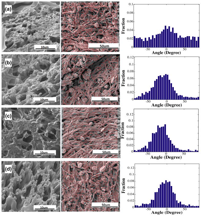
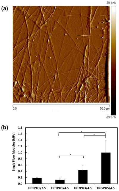
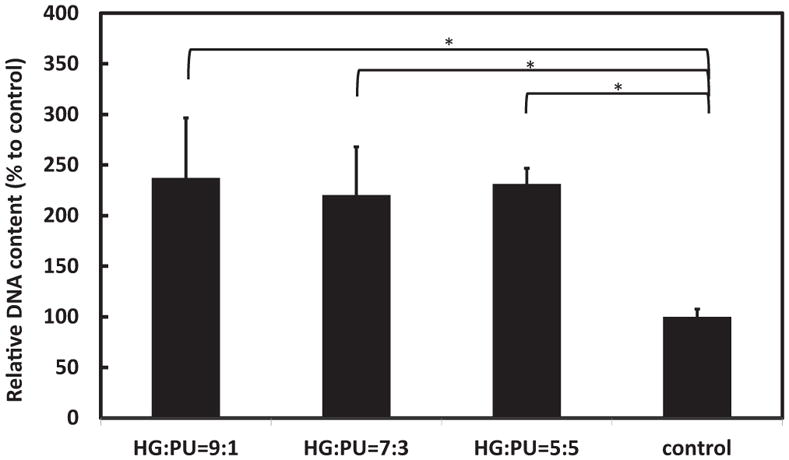
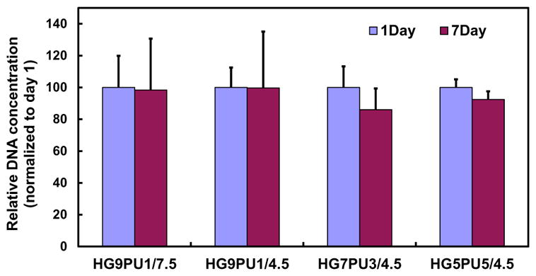
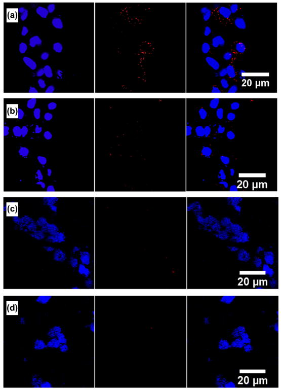
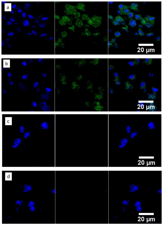
Stem cell therapy has the potential to regenerate heart tissue after myocardial infarction (MI). The regeneration is dependent upon cardiac differentiation of the delivered stem cells. We hypothesized that timing of the stem cell delivery determines the extent of cardiac differentiation as cell differentiation is dependent on matrix properties such as biomechanics, structure and morphology, and these properties in cardiac extracellular matrix (ECM) continuously vary with time after MI. In order to elucidate the relationship between ECM properties and cardiac differentiation, we created an in vitro model based on ECM-mimicking fibers and a type of cardiac progenitor cell, cardiosphere-derived cells (CDCs). A simultaneous fiber electrospinning and cell electrospraying technique was utilized to fabricate constructs. By blending a highly soft hydrogel with a relatively stiff polyurethane and modulating fabrication parameters, tissue constructs with similar cell adhesion property but different global modulus, single fiber modulus, fiber density and fiber alignment were achieved. The CDCs remained alive within the constructs during a 1week culture period. CDC cardiac differentiation was dependent on the scaffold modulus, fiber volume fraction and fiber alignment. Two constructs with relatively low scaffold modulus, ∼50-60kPa, most significantly directed the CDC differentiation into mature cardiomyocytes as evidenced by gene expressions of cardiac troponin T (cTnT), calcium channel (CACNA1c) and cardiac myosin heavy chain (MYH6), and protein expressions of cardiac troponin I (cTnI) and connexin 43 (CX43). Of these two low-modulus constructs, the extent of differentiation was greater for lower fiber alignment and higher fiber volume fraction. These results suggest that cardiac ECM properties may have an effect on cardiac differentiation of delivered stem cells.
Keywords: Cardiac differentiation; Cardiosphere-derived cells; Matrix modulus; Stem cell therapy.
Copyright © 2014 Acta Materialia Inc. Published by Elsevier Ltd. All rights reserved.
Figures


Similar articles
-
Thermosensitive and Highly Flexible Hydrogels Capable of Stimulating Cardiac Differentiation of Cardiosphere-Derived Cells under Static and Dynamic Mechanical Training Conditions.ACS Appl Mater Interfaces. 2016 Jun 29;8(25):15948-57. doi: 10.1021/acsami.6b04932. Epub 2016 Jun 20. ACS Appl Mater Interfaces. 2016. PMID: 27281488 Free PMC article.
-
Differentiation of cardiosphere-derived cells into a mature cardiac lineage using biodegradable poly(N-isopropylacrylamide) hydrogels.Biomaterials. 2011 Apr;32(12):3220-32. doi: 10.1016/j.biomaterials.2011.01.050. Epub 2011 Feb 5. Biomaterials. 2011. PMID: 21296413
-
Production of zebrafish cardiospheres and cardiac progenitor cells in vitro and three-dimensional culture of adult zebrafish cardiac tissue in scaffolds.Biotechnol Bioeng. 2017 Sep;114(9):2142-2148. doi: 10.1002/bit.26331. Epub 2017 Jun 6. Biotechnol Bioeng. 2017. PMID: 28475237
-
Natural ECM as biomaterial for scaffold based cardiac regeneration using adult bone marrow derived stem cells.Stem Cell Rev Rep. 2013 Apr;9(2):158-71. doi: 10.1007/s12015-013-9427-6. Stem Cell Rev Rep. 2013. PMID: 23319217 Review.
-
Biomimetic Extracellular Matrices and Scaffolds Prepared from Cultured Cells.Adv Exp Med Biol. 2018;1078:465-474. doi: 10.1007/978-981-13-0950-2_24. Adv Exp Med Biol. 2018. PMID: 30357638 Review.
Cited by
-
In vitro comparative study of two decellularization protocols in search of an optimal myocardial scaffold for recellularization.Am J Transl Res. 2015 Mar 15;7(3):558-73. eCollection 2015. Am J Transl Res. 2015. PMID: 26045895 Free PMC article.
-
Thermosensitive and Highly Flexible Hydrogels Capable of Stimulating Cardiac Differentiation of Cardiosphere-Derived Cells under Static and Dynamic Mechanical Training Conditions.ACS Appl Mater Interfaces. 2016 Jun 29;8(25):15948-57. doi: 10.1021/acsami.6b04932. Epub 2016 Jun 20. ACS Appl Mater Interfaces. 2016. PMID: 27281488 Free PMC article.
-
Biobased polyurethanes for biomedical applications.Bioact Mater. 2020 Oct 15;6(4):1083-1106. doi: 10.1016/j.bioactmat.2020.10.002. eCollection 2021 Apr. Bioact Mater. 2020. PMID: 33102948 Free PMC article. Review.
-
miRNA-encapsulated abiotic materials and biovectors for cutaneous and oral wound healing: Biogenesis, mechanisms, and delivery nanocarriers.Bioeng Transl Med. 2022 Jun 28;8(1):e10343. doi: 10.1002/btm2.10343. eCollection 2023 Jan. Bioeng Transl Med. 2022. PMID: 36684081 Free PMC article. Review.
-
Synthetic scaffolds for musculoskeletal tissue engineering: cellular responses to fiber parameters.NPJ Regen Med. 2019 Jun 27;4:15. doi: 10.1038/s41536-019-0076-5. eCollection 2019. NPJ Regen Med. 2019. PMID: 31263573 Free PMC article. Review.
References
-
- American Heart Association. Heart Disease and Stroke Statistics—2012 Update. Dallas, TX: American Heart Association; 2012.
-
- Wang F, Guan J. Cellular cardiomyoplasty and cardiac tissue engineering for myocardial therapy. Adv Drug Deliv Rev. 2010;62:784–97. - PubMed
-
- Herrmann JL, Abarbanell AM, Weil BR, Wang Y, Wang M, Tan J, et al. Cell-based therapy for ischemic heart disease: a clinical update. Ann Thorac Surg. 2009;88(5):1714–22. - PubMed
-
- Forrester JS, Makkar RR, Marbán E. Long-term outcome of stem cell therapy for acute myocardial infarction: right results, wrong reasons. J Am Coll Cardiol. 2009;53:2270–2. - PubMed
Publication types
MeSH terms
Substances
Grants and funding
LinkOut - more resources
Full Text Sources
Other Literature Sources
Medical
Research Materials
