Review
. 2014 Jul:41:1-25.
doi: 10.1016/j.preteyeres.2014.04.001. Epub 2014 Apr 21.

Ocular vascular occlusive disorders: natural history of visual outcome

Affiliations
Review

Ocular vascular occlusive disorders: natural history of visual outcome

Sohan Singh Hayreh. Prog Retin Eye Res. 2014 Jul.

Abstract

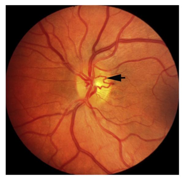
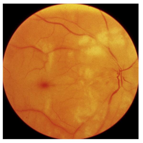
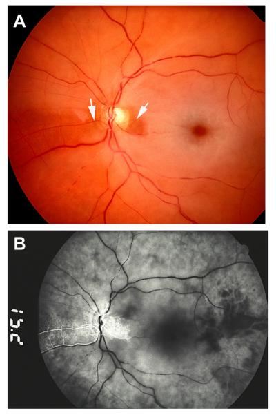
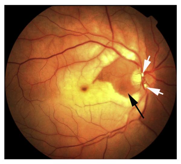
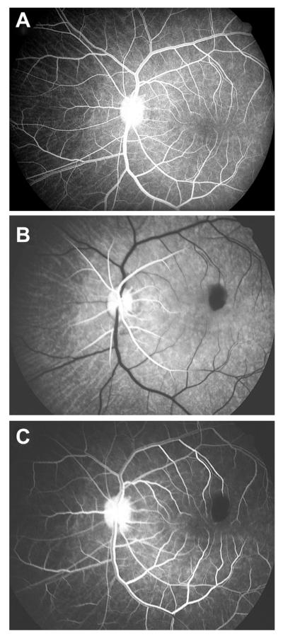
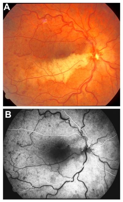
Ocular vascular occlusive disorders collectively constitute the most common cause of visual disability. Before a disease can be managed, it is essential to understand its natural history, so as to be able to assess the likely effectiveness of any intervention. I investigated natural history of visual outcome in prospective studies of 386 eyes with non-arteritic anterior ischemic optic neuropathy (NA-AION), 16 eyes with non-arteritic posterior ischemic optic neuropathy, 697 eyes with central retinal vein occlusion (CRVO), 67 eyes with hemi-CRVO (HCRVO), 216 eyes with branch retinal vein occlusion (BRVO), 260 eyes with central retinal artery occlusion (CRAO), 151 eyes with branch retinal artery occlusion (BRAO) and 61 eyes with cilioretinal artery occlusion (CLRAO). My studies have shown that every one of these disorders consists of multiple distinct clinical sub-categories with different visual findings. When an ocular vascular occlusive disorder is caused by giant cell arteritis, which is an ophthalmic emergency, it would be unethical to do a natural history study of visual outcome in them, because in this case early diagnosis and immediate, intensive high-dose steroid therapy is essential to prevent any further visual loss, not only in the involved eye but also in the fellow, normal eye. In NA-AION in eyes seen ≤2 weeks after the onset, visual acuity (VA) improved in 41% of those with VA 20/70 or worse, and visual field (VF) improved in 26% of those with moderate to severe VF defect. In non-ischemic CRVO eyes with VA 20/70 or worse, VA improved in 47% and in ischemic CRVO in 23%; moderate to severe VF defect improved in 79% in non-ischemic CRVO and in 27% in ischemic CRVO. In HCRVO, overall findings demonstrated that initial VA and VF defect and the final visual outcome were different in non-ischemic from ischemic HCRVO - much better in the former than the latter. In major BRVO, in eyes with initial VA of 20/70 or worse, VA improved in 69%, and moderate to severe VF defect improved in 52%. In macular BRVO with 20/70 or worse initial VA, it improved in 53%, and initial minimal-mild VF defect was stable or improved in 85%. In various types of CRAO there are significant differences in both initial and final VA and VF defects. In CRAO eyes seen within 7 days of onset and initial VA of counting fingers or worse, VA improved in 82% with transient non-arteritic CRAO, 67% with non-arteritic CRAO with cilioretinal artery sparing, 22% with non-arteritic CRAO. Central VF improved in 39% of transient non-arteritic CRAO, 25% of non-arteritic CRAO with cilioretinal artery sparing and 21% of non-arteritic CRAO. Peripheral VF improved in non-arteritic CRAO in 39% and in transient non-arteritic CRAO in 39%. In transient CRAO, finally peripheral VFs were normal in 93%. In non-arteritic CRAO eyes initially 22% had normal peripheral VF and in the rest it improved in 39%. Final VA of 20/40 or better was seen in 89% of permanent BRAO, and in 100% of transient BRAO and non-arteritic CLRAO. In permanent BRAO eyes, among those seen within 7 days of onset, central VF defect improved in 47% and peripheral VF in 52%, and in transient BRAO central and peripheral VFs were normal at follow-up. My studies showed that AION, CRVO, BRVO, CRAO and BRAO, each consist of multiple distinct clinical sub-categories with different visual outcome. Contrary to the prevalent impression, these studies on the natural history of visual outcome have shown that there is a statistically significant spontaneous visual improvement in each category. The factors which influence the visual outcome in various ocular vascular occlusive disorders are discussed.

Keywords: Branch retinal vein occlusion; Central retinal artery occlusion; Central retinal vein occlusion; Non-arteritic anterior ischemic optic neuropathy.


Figures

Fig. 1
Fig. 1
Fundus photograph of a resolved non-ischemic CRVO in the right eye. It shows retinociliary collaterals on the optic disc, macular retinal pigmentary degeneration and engorged tortuous retinal veins.
Fig. 2
Fig. 2
Fluorescein fundus angiogram of left eye, soon after onset of ischemic CRVO, during the arteriovenous phase (32 s after the injection of fluorescein), shows complete filing of the retinal vasculature.
Fig. 3
Fig. 3
Schematic representation of the blood vessels in the optic nerve. (Modified from Hayreh, 1974a,; 78: OP240-OP254.) Abbreviations: A = arachnoid; C = choroid; CAR and CRA = central retinal artery; Col. Br. = Collateral branches; CRV = central retinal vein; D = dura; LC = lamina cribrosa; ON = optic nerve; PCA = posterior ciliary artery; PR = prelaminar region; R = retina; S = sclera; SAS = subarachnoid space.
Fig. 4
Fig. 4
Cast of the central retinal vein shows its entire course from the optic disc to its exit from the optic nerve sheath. Note the presence of a large number of prominent tributaries within the optic nerve and none in the optic nerve head region in this specimen. (Reproduced from Hayreh et al., 2011a, 118: 119–33.) Abbreviations: CRV = Central retinal vein; ON = optic nerve; ONH = optic nerve head.
Fig. 5
Fig. 5
Schematic representation of two trunks of central retinal vein in the optic nerve. Abbreviations: A = arachnoid; C = choroid; CRA = central artery of retina; CRV = central retinal vein; D = dural sheath; LC = lamina cribrosa; OD = optic disc; ON = optic nerve; PR = prelaminar region; SAS = subarachnoid space; R = retina; S = sclera; P = pia mater. (Reproduced from Hayreh and Hayreh, 1980; 98: 1600–1609.)
Fig. 6
Fig. 6
Diagrammatic reconstruction from serial sections (10-μm thick) of anterior part of optic nerve. (A) It shows intraneural course of central retinal vessels. Note duplicate trunks the central retinal vein anteriorly. (B) One of the transverse sections of the specimen shows, in the center of the optic nerve, two trunks of the central retinal vein, with the central retinal artery (filled with Prussian blue) interposed between the two veins. CRA = central retinal artery; CRV = central retinal vein; ON, optic nerve. (Reproduced from Hayreh SS. Master of surgery thesis. Panjab University, India 1958.)
Fig. 7
Fig. 7
Right eye of a 67-year-old man with non-ischemic HCRVO involving lower half of retina. (A) Fundus photograph during the acute phase. (Two arrows show 2 trunks of central retinal vein). (B, C) Fluorescein fundus angiograms during: (B) during arteriovenous phase shows delayed filling of the occluded vein, and (C) late phase. (D) After resolution of retinopathy shows macular pigmentary degeneration and venous collaterals on the optic disc. (A, B and C reproduced from Hayreh and Hayreh, 1980; 98: 1600–1609.)
Fig. 8
Fig. 8
Left eye of a 67-year-old man with ischemic HCRVO involving lower half of retina, 18 months after the onset. (A) Composite fundus photograph shows optic disc and retinal neovascularization. (B) Fluorescein fundus angiogram during early arteriovenous phase, and (C) during the venous phase shows retinal capillary non-perfusion in the involved retina and fluorescein leak from the neovascularization. (Reproduced from Hayreh and Hayreh, 1980; 98: 1600–1609.)
Fig. 9
Fig. 9
Left eye of an 83-year woman with non-ischemic HCRVO, involving upper half of the retina and entire macular region.
Fig. 10
Fig. 10
Left eye of a 68-year-old man with ischemic HCRVO of 3½ months’ duration, involving upper half of retina, with iris neovascularization, and neovascular glaucoma. Two weeks later, optic disc neovascularization developed. (A) Composite fundus photograph. (B) Fluorescein fundus angiogram during arteriovenous phase shows retinal capillary non-perfusion in the involved retina. (Reproduced from Hayreh and Hayreh, 1980, 98: 1600–1609.)
Fig. 11
Fig. 11
Right eye of a 39-year-old man with non-ischemic HCRVO involving superior temporal sector only.
Fig. 12
Fig. 12
Left eye of a 66-year old man with non-ischemic HCRVO involving inferior temporal region only, 5 months after the onset, and shows venous collateral on the optic disc connecting the two trunks of the central retinal vein (arrow).
Fig. 13
Fig. 13
Left eye of a 48-year woman with non-ischemic HCRVO involving inferior half of the retina 14 months after the onset shows venous collateral on the optic disc connecting the two trunks of the central retinal vein (arrow).
Fig. 14
Fig. 14
Fundus photograph of the right eye with CRAO. It shows retinal infarction, cherry red spot, optic disc edema and narrow retinal arterioles.
Fig. 15
Fig. 15
Left eye of a 77-year old woman with CRAO and cilioretinal artery sparing. (A) Fundus photograph shows central retinal macular ischemic opacity, cherry red spot and box-carring (cattle trucking). Arrows indicate two patent cilioretinal arteries. (B) Fluorescein fundus angiogram shows filling of the cilioretinal arteries and the choroid, but no filling of the central retinal artery circulation 15.2 s after injection of the dye.
Fig. 16
Fig. 16
Right eye of a 35-year old man with CRAO and cilioretinal artery sparing. There are two emboli (white arrows) situated in two branches of the central retinal artery on the optic disc. Black arrow indicates the area supplied by the patent cilioretinal artery. Reproduced by courtesy of Dr. Patel from India.
Fig. 17
Fig. 17
An example of a common trunk of origin of central retinal artery and posterior ciliary artery (PCA) from the ophthalmic artery, as seen from below. Reproduced from Hayreh, 2009; 28: 34–62. CAR = Central retinal artery, LPCA = Lateral PCA, MPCA = Medial PCA, OA = Ophthalmic artery, ON = Optic nerve. PPS = Point of penetration into the sheath by CAR, * = Common trunk of origin of CRA and medial PCA.
Fig. 18
Fig. 18
Right eye of a 69-year old man with giant cell arteritis, with arteritic anterior ischemic optic neuropathy and CRAO. (A) Fundus photograph shows chalky-white optic disc edema, some mild box-carring (cattle-trucking) in the retinal vessels and cherry red spot with perifoveolar ischemic retinal opacity. (B) Fluorescein fundus angiogram during the transit of the dye shows late and slow filling of the central retinal artery and of a few patches of the choroid. (C) Fluorescein fundus angiogram during the late phase shows box-carring (cattle-trucking) in the retinal vessels, with almost no disc staining, partial filling of the choroid.
Fig. 19
Fig. 19
Right eye of a 69-year old man; one day old transient CRAO. (A) Fundus photograph shows central macular retinal infarct with cherry red spot and normal retinal vessels. (B) Fluorescein fundus angiogram that day during the retinal arteriovenous phase shows normal retinal vascular bed filling, with poor filling of the central foveal retinal capillaries. (C) Visual fields plotted with a Goldmann perimeter, show normal peripheral field with I-4e, island field with I-2e, and a large absolute central scotoma.
Fig. 20
Fig. 20
Left eye of a 30-year old man with transient CRAO. (A) Fundus photograph shows ischemic retinal opacity in the macular region, with a few punctate hemorrhages, normal retinal vessels, and tiny patent cilioretinal artery (Arrow). (B) Fluorescein fundus angiogram 30 s after injection of the dye shows filling of the retinal vessels but no filling of the retinal vessels in the central macular region.
Fig. 21
Fig. 21
Fluorescein fundus angiograms of the left eye of a rhesus monkey, (A) before and (B,C) immediately after experimental cutting of the central retinal artery at its site of entry into the optic nerve. (A) Before cutting the artery, normal angiogram 14 s after injection of the dye, shows normal retinal vascular filling during the late arteriovenous phase. (B) After cutting the artery, angiogram 14 s after injection of the dye, shows start of filling of the retinal arterioles only up to a short distance away the disc. (C) After cutting the artery, angiogram 52 s after the injection of the dye, showing retinal vascular filling during the late arteriovenous phase, corresponding to the phase seen in (A) above. Reproduced from Hayreh, 2005; 24: 493–519.
Fig. 22
Fig. 22
Transverse section of a human optic nerve at the level of entry of the central retinal artery into the optic nerve sheath. It shows the central retinal artery lying in the dural sheath. CRA = central retinal artery; ON = Optic nerve. (Reproduced from Hayreh SS. Master of surgery thesis. Panjab University, India 1958.)
Fig. 23
Fig. 23
Right fundus photograph of a 71-year old woman with cilioretinal artery occlusion (Arrow).
Fig. 24
Fig. 24
Left eye with arteritic anterior ischemic optic neuropathy and cilioretinal artery occlusion in 63-year woman with giant cell arteritis. (A) Fundus photograph shows chalky-white optic disc edema and a patch of retinal opacity in the distribution of the cilioretinal artery occlusion (Arrow). (B) Fluorescein fundus angiogram shows normal filling of the central retinal artery and of the choroid supplied by the lateral posterior ciliary artery, but no filling of the choroid and optic disc supplied by the medial posterior ciliary artery as well as of the cilioretinal artery (arrow). (Reproduced from Hayreh, 1981; 38: 675–678.)
Fig. 25
Fig. 25
Right eye of a 32-year old man with cilioretinal artery occlusion and non-ischemic central retinal vein occlusion. (A) Fundus photograph shows retinal infarct in the region of the occluded cilioretinal artery, with engorged retinal veins, sparse retinal hemorrhages and retinociliary collaterals on the optic disc. (Reproduced from Hayreh et al., 2008; 28: 581–594.) (B) Fluorescein fundus angiogram during the retinal arterial phase shows non-filling of the retina supplied by the occluded cilioretinal artery.

References

    1. Arnold AC, Hepler RS. Natural history of nonarteritic anterior ischemic optic neuropathy. J. Neuroophthalmol. 1994;14:66–69. - PubMed
    1. Brown GC, Magargal LE. Central retinal artery obstruction and visual acuity. Ophthalmology. 1982;89:14–19. - PubMed
    1. Brown GC, Moffat K, Cruess A, Magargal LE, Goldberg RE. Cilioretinal artery obstruction. Retina. 1983;3:182–187. - PubMed
    1. Chen JC, Klein ML, Watzke RC, Handelman IL, Robertson JF. Natural course of perfused central retinal vein occlusion. Can. J. Ophthalmol. 1995;30:21–24. - PubMed
    1. Chopdar A. Hemi-central retinal vein occlusion. Pathogenesis, clinical features, natural history and incidence of dual trunk central retinal vein. Trans. Ophthalmol. Soc. U. K. 1982;102:241–248. - PubMed

Publication types

MeSH terms