Review
. 2014 Jun:43:183-90.
doi: 10.1016/j.neubiorev.2014.04.009. Epub 2014 Apr 23.

Developmental studies of the hippocampus and hippocampal-dependent behaviors: insights from interdisciplinary studies and tips for new investigators

Affiliations
Review

Developmental studies of the hippocampus and hippocampal-dependent behaviors: insights from interdisciplinary studies and tips for new investigators

Sarah H Albani et al. Neurosci Biobehav Rev. 2014 Jun.

Abstract

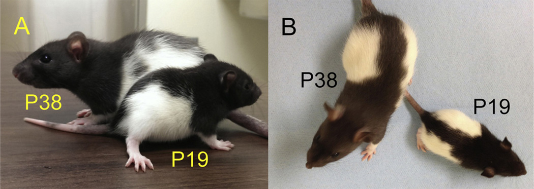
The hippocampus is not fully developed at birth and, with respect to spatial cognition, only begins to show signs of adult-like function at three postnatal weeks in rodents. Studying the developmental period spanning roughly two to four weeks of age permits an understanding of the neural framework necessary for the emergence of spatial navigation and, quite possibly, human episodic memory. However, due to developmental factors, behavior data collection and interpretation can be severely compromised if inappropriate designs are applied. As such, we propose methodological considerations for the behavioral assessment of hippocampal function in developing rats that take into account animal size, growth rate, and sensory and motor ability. We further summarize recent key interdisciplinary studies that are beginning to unravel the molecular machinery and physiological alterations responsible for hippocampal maturation. In general, hippocampal development is a protracted process during which unique contributions to spatial cognition and complex recognition memory come "on line" at different postnatal ages creating a unique situation for elucidating the neural bases of specific components of higher cognitive abilities.

Keywords: AMPAR; Behavioral testing; Environment; Hippocampus; Juvenile; Learning and memory; NMDAR; Novelty; Place field; Postnatal development; Spatial navigation; Y-maze.


Figures

Fig. 1
Fig. 1
Long–Evans rats at P19 and P38. Both height (A) and length (B) differ dramatically between these age groups. Behavioral testing conditions must account for differences in size and step length in relation to the animal’s age.
Fig. 2
Fig. 2
Y-maze environment for juvenile (A) and adult (B) rodents. Maze dimensions are reduced to accommodate the smaller juvenile. Visual cues for juvenile testing should be larger and separated by a greater distance than their adult counterparts to allow for developmental differences in visual acuity.
Fig. 3
Fig. 3
Object–place (A), object–context (B), and object–place–context (C) recognition tasks as described by Langston and Wood (2010). The cylinder and cube represent two different objects. (A) In the sample phase of the object–place task, two different objects (green cube and orange cylinder) are presented to the rat. In the test phase, two objects of the same shape are presented in the same context, such that one object (the circled cube) is in a novel place. (B) In the sample 1 phase of the object–context task, two objects of the same shape are presented in spatial context 1. In the sample 2 phase, two copies of a different object are presented in spatial context 2 (denoted by the blue platform). The test condition maintains context 2 and swaps one object from context 2 with one object from context 1, such that one object (the circled cube) is in a novel object–context configuration. (C) In the sample 1 phase of the object–place–context recognition task, two different objects are presented in context 1. In the sample 2 phase, the context is changed and the object locations are swapped. The test condition returns to the original context and presents a copy of one object from the original context with one object from the second context in the place of the other object, such that one object (the circled cube) is in a novel object–place–context configuration. Typically, there is a two-minute latency between phases of each recognition experiment. (For interpretation of the references to color in this figure legend, the reader is referred to the web version of the article.)
Fig. 4
Fig. 4
Single units (individual action potential discharge events) and LFPs reflecting population-level events can be measured in real time during arena exploration. (A)Typically, tetrodes are implanted so that the tips of the electrodes reside in the cell body layer of hippocampal area CA1. (B) Signals are high-pass filtered to observe the pattern of responses that individual action potentials produce on each electrode, permitting isolation of multiple distinct units (X and Y) by each tetrode. (C) One or more electrodes can be used to record population LFPs. Power scores can be calculated to determine the amount that any frequency or range of frequencies is represented in the population trace. Oscillatory rhythms in the range of 6–12 (theta) and 45–120 Hz (gamma) are often analyzed with respect to behavior of the animal. The example LFP is filtered at 3–140 Hz. Scale bar is one second. (D) The exploration path of an animal in a circular arena (left) shown with the heat map of discharge frequency for a single unit (right) reveals the place field of a single neuron. Firing rate increases when the animal occupies the place field and different units have different, but sometimes overlapping place fields. (E) During prospective encoding, neurons with established place fields (indicated by the colored dots on the linear maze and waveforms at top) fire transiently in advance of the animal’s movement and sweep from one place field (Neuron A) to the next (Neuron B) toward the animal’s destination (X) (Johnson and Redish, 2007). In a two-dimensional maze, neurons with established place fields (indicated by the colored dots) show prospective encoding by firing sequentially (red, blue, then green) toward the animal’s destination (X). Neurons sweep toward a home location (X) independent of the animal’s previous outbound path (Pfeiffer and Foster, 2013). (For interpretation of the references to color in this figure legend, the reader is referred to the web version of the article.)
Fig. 5
Fig. 5
Developmental modifications to AMPA receptors and NMDA receptors at excitatory synapses in the hippocampus. (A) During the third postnatal week, NMD A receptors with GluN2B subunits (blue) are replaced by NMDA receptors with GluN2A subunits (green). (B) From P17 to P24, expression of the AMPA receptor subunit, GluA1 (red), decreases while expression of GluA3 (yellow) and transmembrane AMPA receptor regulatory protein TARP (brown) increase. Synapse associated protein (SAP102) and postsynaptic density protein (PSD95) are anchoring proteins (orange) for AMPA and NMDA receptors at glutamatergic synapses (Elias et al., 2008). TARP regulates synaptic anchoring and AMPA receptor channel dynamics (Jackson and Nicoll, 2011). (For interpretation of the references to color in this figure legend, the reader is referred to the web version of the article.)
Fig. 6
Fig. 6
Expression of chimeric GluN2 subunits allows for separation of the roles of NMDA receptor-dependent calcium-conductance and intracellular protein–protein signaling in the maturation of glutamatergic synapses and spatial navigation. (A) A shift from predominantly GluN2B (blue) to GluN2A subunits (green) is associated with mature hippocampal function in wildtype animals. In the ABc chimera, the GluN2A regulatory domains for calcium conductance (amino and transmembrane, TM) are fused to the Glun2B intracellular protein-protein signaling domains (carboxy terminus). (B) Animals expressing ABc (blue and green striped bars) display precocious spatial navigation ability at P17–19 as compared to wildtype littermates (blue bar). This suggests that incorporation of GluN2A-type calcium conductance domains (red box in subunit illustrations), more so than intracellular protein–protein signaling, permits the late postnatal emergence of hippocampus-dependent behavior (red box in bar graph). (For interpretation of the references to color in this figure legend, the reader is referred to the web version of the article.) Adapted from Sanders et al. (2013).

Similar articles

Cited by

References

    1. Aadland J, Beatty WW, Maki RH. Spatial memory of children and adults assessed in the radial maze. Dev. Psychobiol. 1985;18:163–172. http://dx.doi.org/10.1002/dev.420180208. - DOI - PubMed
    1. Ainge JA, Tamosiunaite M, Woergoetter F, Dudchenko PA. Hippocampal CA1 place cells encode intended destination on a maze with multiple choice points. J. Neurosci. 2007;27:9769–9779. http://dx.doi.org/10.1523/JNEUROSCI.2011-07.2007. - DOI - PMC - PubMed
    1. Ainge JA, Tamosiunaite M, Worgotter F, Dudchenko PA. Hippocampal place cells encode intended destination, and not a discriminative stimulus, in a conditional T-maze task. Hippocampus. 2012;22:534–543. http://dx.doi.org/10.1002/hipo.20919. - DOI - PubMed
    1. Arai AC, Kessler M. Pharmacology of ampakine modulators: from AMPA receptors to synapses and behavior. Curr. Drug Targets. 2007;8:583–602. http://dx.doi.org/10.2174/138945007780618490. - DOI - PubMed
    1. Barker GR, Warburton EC. When is the hippocampus involved in recognition memory? J. Neurosci. 2011;31:10721–10731. http://dx.doi.org/10.1523/JNEUROSCI.6413-10.2011. - DOI - PMC - PubMed

Publication types