

Imaging outcomes for trials of remyelination in multiple sclerosis
- PMID: 24769473
- PMCID: PMC4335693
- DOI: 10.1136/jnnp-2014-307650
Imaging outcomes for trials of remyelination in multiple sclerosis
Abstract
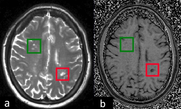
Trials of potential neuroreparative agents are becoming more important in the spectrum of multiple sclerosis research. Appropriate imaging outcomes are required that are feasible from a time and practicality point of view, as well as being sensitive and specific to myelin, while also being reproducible and clinically meaningful. Conventional MRI sequences have limited specificity for myelination. We evaluate the imaging modalities which are potentially more specific to myelin content in vivo, such as magnetisation transfer ratio (MTR), restricted proton fraction f (from quantitative magnetisation transfer measurements), myelin water fraction and diffusion tensor imaging (DTI) metrics, in addition to positron emission tomography (PET) imaging. Although most imaging applications to date have focused on the brain, we also consider measures with the potential to detect remyelination in the spinal cord and in the optic nerve. At present, MTR and DTI measures probably offer the most realistic and feasible outcome measures for such trials, especially in the brain. However, no one measure currently demonstrates sufficiently high sensitivity or specificity to myelin, or correlation with clinical features, and it should be useful to employ more than one outcome to maximise understanding and interpretation of findings with these sequences. PET may be less feasible for current and near-future trials, but is a promising technique because of its specificity. In the optic nerve, visual evoked potentials can indicate demyelination and should be correlated with an imaging outcome (such as optic nerve MTR), as well as clinical measures.
Keywords: MRI; Multiple Sclerosis.
Published by the BMJ Publishing Group Limited. For permission to use (where not already granted under a licence) please go to http://group.bmj.com/group/rights-licensing/permissions.
Figures
References
-
- Barkhof F, Calabresi PA, Miller DH, et al. Imaging outcomes for neuroprotection and repair in multiple sclerosis trials. Nat Rev Neurol 2009;5:256–66. - PubMed
-
- Bitsch A, Kuhlmann T, Stadelmann C, et al. A Longitudinal MRI Study of Histopathologically Defined Hypointense Multiple Sclerosis Lesions. Ann Neurol 2001;49:793–6. - PubMed
-
- Van Walderveen MA, Kamphorst W, Scheltens P, et al. Histopathologic correlate of hypointense lesions on T1-weighted spin-echo MRI in multiple sclerosis. Neurology 1998;50:1282–8 http://www.ncbi.nlm.nih.gov/pubmed/9595975 - PubMed
-
- Vavasour IM, Laule C, Li DKB, et al. Is the magnetization transfer ratio a marker for myelin in multiple sclerosis? J Magn Reson Imaging 2011;33:713–8. - PubMed
-
- Schmierer K, Scaravilli F, Altmann DR, et al. Magnetization transfer ratio and myelin in postmortem multiple sclerosis brain. Ann Neurol 2004;56:407–15. - PubMed
Publication types
MeSH terms
LinkOut - more resources
Full Text Sources
Other Literature Sources
Medical