. 2014 Apr;26(4):1497-1511.
doi: 10.1105/tpc.114.124586. Epub 2014 Apr 25.

A Receptor-Like Kinase Mediates Ammonium Homeostasis and Is Important for the Polar Growth of Root Hairs in Arabidopsis

Affiliations

A Receptor-Like Kinase Mediates Ammonium Homeostasis and Is Important for the Polar Growth of Root Hairs in Arabidopsis

Ling Bai et al. Plant Cell. 2014 Apr.

Abstract

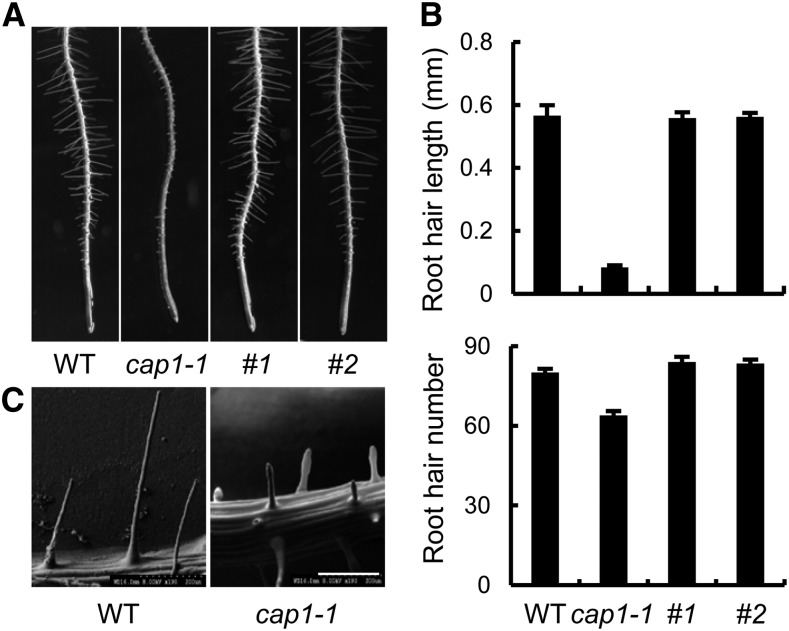
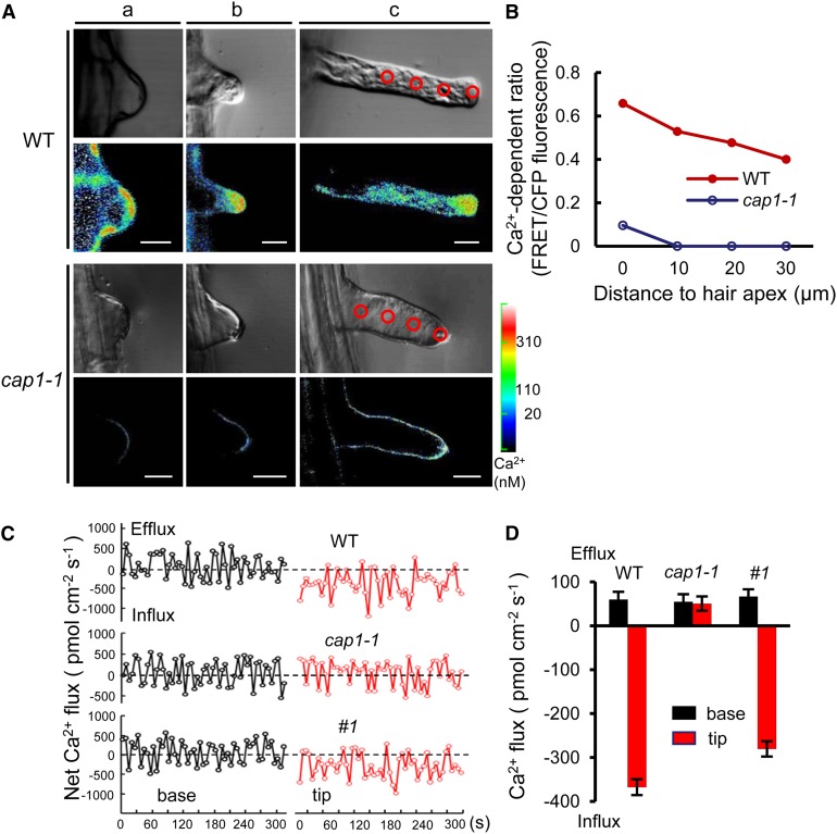
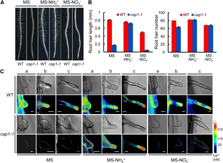
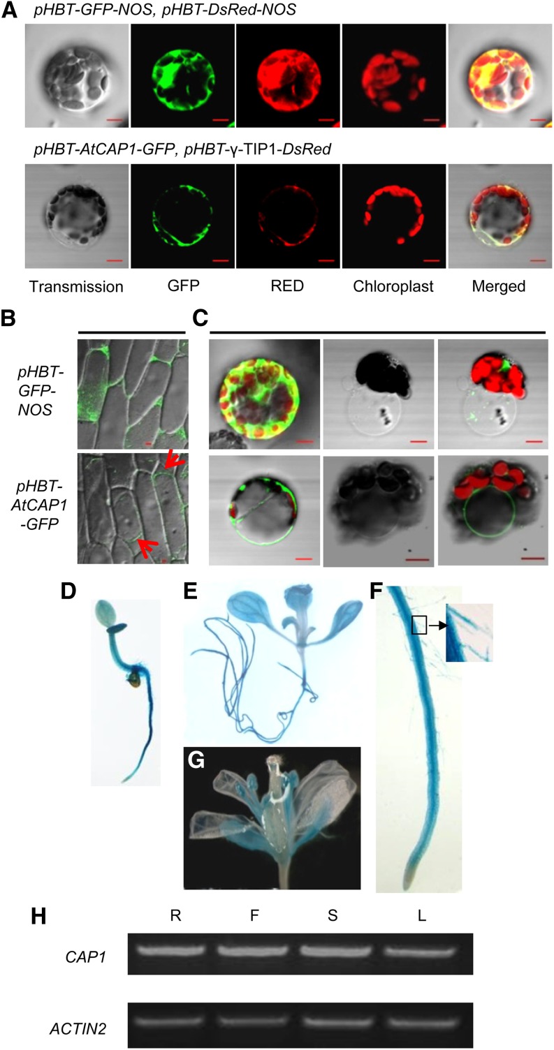
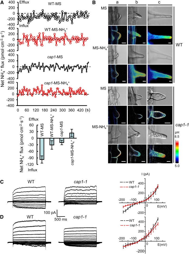
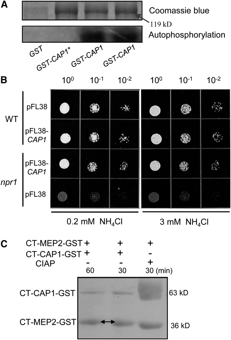
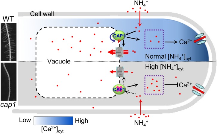
Ammonium (NH4+) is both a necessary nutrient and an important signal in plants, but can be toxic in excess. Ammonium sensing and regulatory mechanisms in plant cells have not been fully elucidated. To decipher the complex network of NH4+ signaling, we analyzed [Ca2+]cyt-associated protein kinase (CAP) genes, which encode signaling components that undergo marked changes in transcription levels in response to various stressors. We demonstrated that CAP1, a tonoplast-localized receptor-like kinase, regulates root hair tip growth by maintaining cytoplasmic Ca2+ gradients. A CAP1 knockout mutant (cap1-1) produced elevated levels of cytoplasmic NH4+. Furthermore, root hair growth of cap1-1 was inhibited on Murashige and Skoog medium, but NH4+ depletion reestablished the Ca2+ gradient necessary for normal growth. The lower net NH4+ influx across the vacuolar membrane and relatively alkaline cytosolic pH of cap1-1 root hairs implied that mutation of CAP1 increased NH4+ accumulation in the cytoplasm. Furthermore, CAP1 functionally complemented the npr1 (nitrogen permease reactivator protein) kinase yeast mutant, which is defective in high-affinity NH4+ uptake via MEP2 (methylammonium permease 2), distinguishing CAP1 as a cytosolic modulator of NH4+ levels that participates in NH4+ homeostasis-regulated root hair growth by modulating tip-focused cytoplasmic Ca2+ gradients.


Figures

Figure 1.
Figure 1.
Mutation of CAP1 Impairs Root Hair Growth in Arabidopsis. (A) Root hair growth of the wild type (WT), mutant (cap1-1), and complementation lines (#1 and #2). Seedlings were grown on vertical 1.2% agar MS medium for 7 d. (B) Comparison of root hair lengths and numbers between the wild type, cap1-1, and complementation lines. Root hair lengths were measured in 7-d-old seedlings 5 mm from the primary root tips. Data bars represent means ± se of root hair lengths from triplicate experiments (wild type and cap1-1, n = 100; complementation lines, n = 60). (C) Scanning electron micrographs of wild-type and cap1-1 root hairs. Bar = 200 µm.
Figure 2.
Figure 2.
Tip-Focused Ca2+ Gradients and Net Fluxes of Ca2+ in Root Hair Tips Disappeared in cap1-1 Mutants. (A) Tip-focused Ca2+ gradients in the cytoplasm of Arabidopsis wild-type and cap1-1 root hairs expressing the Ca2+ sensor YC3.6 at different developmental stages (a, initiation phase; b, transition phase; and c, tip growth phase). Bright-field and fluorescence ratio images of Ca2+ in root hairs of the wild type and cap1-1 were obtained as described by Monshausen et al. (2008). Cytosolic Ca2+ levels were pseudo-color-coded according to the scale. Representative images of more than 10 measurements from three separate experiments per genotype are presented. Bars = 10 µm. (B) Quantitative analysis of cytosolic Ca2+ levels in a representative growing root hair. Relative Ca2+ concentrations were measured in 10-µm2 regions of interest along lengths of the root hairs in (A), as indicated with circles in the bright-field images. An increase in the fluorescence resonance energy transfer/cyan fluorescent protein ratio based on the Ca2+ sensor YC3.6 reflects an increase in cytoplasmic Ca2+ level. (C) Ca2+ flux profiles of root hairs in the wild type, cap1-1, and cap1-1/CAP1. Ion-selective vibration microelectrode recordings of Ca2+ fluxes at the surfaces of root hairs of 7-d-old seedlings were made. Graphs show data from positions corresponding to the base (left) and tip (right) of root hairs (Supplemental Figure 4). Trace is a recording of a typical experimental plot illustrating Ca2+ influx and efflux in a root hair (inwards, negative; outwards, positive). (D) Mean fluxes of Ca2+ in root hairs. Bars represent means ± se (n = 5).
Figure 3.
Figure 3.
NH4+ Deprivation Restored cap1-1 Root Hair Growth and Tip-Focused Ca2+ Gradient. (A) The absence of NH4+, but not NO3, restored root hair growth in cap1-1. Seedlings of the wild type and cap1-1 grown for 7 d on MS, MS lacking NH4+ (MS-NH4+), and MS lacking NO3 (MS-NO3). Root hairs of cap1-1 were only restored in NH4+-free medium. (B) Comparison of root hair lengths and numbers between the wild type and cap1-1 on MS, MS-NH4+, and MS-NO3 media. Data were collected from seedlings grown on MS (n = 100), NH4+-free (n = 90), and NO3 deprivation (n = 60) media from three separate experiments as described in Figure 1B. Error bars represent means ± se. (C) The tip-focused Ca2+ gradient was reestablished in YC3.6-transformed cap1-1 on NH4+-free but not on NO3 deprivation media. The [Ca2+]cyt of root hairs was measured as described in Figure 2A (a, initiation phase; b, transition phase; and c, tip growth phase). Bars = 10 µm.
Figure 4.
Figure 4.
CAP1 Localizes to the Tonoplast and Is Expressed in Root Hairs. (A) to (C) Confocal analysis of the subcellular localization of CAP1. Bars = 10 µm. (A) CAP1-fused GFP was transiently coexpressed with the vacuolar membrane marker γ-TIP1 in protoplasts. Images showed colocalization of CAP1 and γ-TIP1 (bottom panels). Empty vector pHBT-GFP-NOS expressed in protoplasts was a control (top panels). (B) Transient expression in onion epidermal cells shows fluorescence of CAP1 in the vacuolar membrane. Arrows point to the vacuole. (C) Fluorescence images of GFP-transformed protoplast (top panels) and GFP-fused, CAP1-transformed protoplast (bottom panels). The same protoplast before (first image) and immediately after bursting (second two images) is shown. (D) to (G) CAP1 expression pattern. GUS staining of transgenic plants shows CAP1 expressed in roots ([D] to [F]), leaves (E), and flowers (G). Inset image shows root hairs in (F). (H) RT-PCR indicated that CAP1 was expressed in roots (R), flowers (F), inflorescence stems (S), and young leaves (L).
Figure 5.
Figure 5.
CAP1 Modulates Transportation of Ammonium from the Cytoplasm to the Vacuole. (A) Changes in NH4+ net fluxes of root hair vacuoles (inwards, negative; outwards, positive). Points are data collected every 6 s. Typical net flux traces are shown in the top panel. The NH4+ net fluxes are averaged from MS (n = 17 for wild type, n = 15 for cap1) and MS-NH4+ media (n = 7 for wild type, n = 9 for cap1) and plotted in the bottom panel. Error bars represent means ± se. (B) Visualizing pHc in root hairs in stable transgenic wild-type and cap1-1 plants with phGFP using ratiometric pH-sensitive GFP. phGFP accumulates in the peripheral cytoplasm regions near the plasma membrane in cap1-1 seedling’s root hair cells grown on MS medium. pH levels were pseudo-color-coded according to the calibrated 410-nm/470-nm ratio image of the same root hair (Supplemental Figure 6). The different phases of root hair development for the wild type and cap1-1 are shown: a, the initial phase; b, transition phase; c, tip growth phase. Bars = 10 µm. (C) Whole-cell voltage-activated currents in root hair protoplasts of the wild type and cap1-1 mutants. Typical time-dependent currents recorded in root hair cell protoplasts of the wild type (left) and cap1-1 (middle) and NH4+ current-voltage relationships for the wild type (n = 4) and cap1-1 (n = 6) (I-V curve, right) are shown. I-V curves show means ± se. (D) Effects of NH4+ on whole-cell currents of wild-type and cap1-1 root hairs. The data of NH4+ application to root hair cells by the addition of 150 mM NH4+ to a pipette solution were recorded in the whole-cell patch-clamp configuration (wild type, n = 7; cap1-1, n = 4).
Figure 6.
Figure 6.
CAP1 Can Be Autophosphorylated and Complements the Activity of NPR1 Kinase in Yeast. (A) In vitro phosphorylation of CAP1 kinase activity. Recombinant protein fused with GST was used in the kinase assay. Boiled CAP1 protein (GST-CAP1*) was a negative control. Autophosphorylation was detected after protein gel electrophoresis and phosphor imaging. (B) Growth of yeast strains on solid minimal medium containing different concentrations of NH4+ as the sole nitrogen source. Strains 23344c (ura3, wild type) and 21994b (npr1-1, ura3) transformed with plasmid pFL38 and pFL38-CAP1 were spotted at 100-, 10−1-, and 10−2-fold dilutions on YNB medium supplemented with 0.2 and 3 mM NH4Cl and incubated for 4 d at 29°C. (C) In vitro kinase assay for MEP2 phosphorylation by CAP1. Recombinant C-terminal fragments of CAP1 (CT-CAP1-GST) and MEP2 (CT-MEP2-GST) were cultured in kinase buffer, and phosphorylated MEP2 exhibited slower mobility (arrows). MEP2 treated with CIAP (alkaline phosphatase) was the control. The phosphorylation reactions were stopped by boiling for 5 min, and the proteins were separated by SDS-PAGE.
Figure 7.
Figure 7.
Model Showing the Putative Regulation Pathway of NH4+ Homeostasis Mediated by CAP1 in Root Hairs. In the wild type (in blue), CAP1 in the vacuolar membrane senses cytosolic NH4+ levels and phosphorylates an unknown target, such as tonoplast intrinsic protein, resulting in compartmentalization of NH4+ in the vacuole and the maintenance of normal NH4+ levels in the cytoplasm. Normal NH4+ homeostasis is necessary for the establishment of the [Ca2+]cyt gradient in the polar growth of root hairs. When CAP1 was rendered deficient in the mutant (in gray), inward NH4+ flux across the vacuole membrane significantly decreased and excess NH4+ accumulated in the cytoplasm, causing a loss of calcium gradient (black line with block end) and eventual cessation of root hair growth. The cytosolic Ca2+ gradient in the wild type is indicated by the scale below. No gradient was established in cap1 mutants. Red dots represent ammonium ions. Red lines with arrows indicate ion influx.

References

    1. Alonso J.M., et al. (2003). Genome-wide insertional mutagenesis of Arabidopsis thaliana. Science 301: 653–657 - PubMed
    1. Babourina O., Voltchanskii K., McGann B., Newman I., Rengel Z. (2007). Nitrate supply affects ammonium transport in canola roots. J. Exp. Bot. 58: 651–658 - PubMed
    1. Bei Q., Luan S. (1998). Functional expression and characterization of a plant K+ channel gene in a plant cell model. Plant J. 13: 857–865 - PubMed
    1. Benfey P.N., Bennett M., Schiefelbein J. (2010). Getting to the root of plant biology: impact of the Arabidopsis genome sequence on root research. Plant J. 61: 992–1000 - PMC - PubMed
    1. Bernhardt C., Lee M.M., Gonzalez A., Zhang F., Lloyd A., Schiefelbein J. (2003). The bHLH genes GLABRA3 (GL3) and ENHANCER OF GLABRA3 (EGL3) specify epidermal cell fate in the Arabidopsis root. Development 130: 6431–6439 - PubMed