. 2014 Jul;196(13):2455-71.
doi: 10.1128/JB.01768-14. Epub 2014 Apr 25.

Analysis of surface-exposed outer membrane proteins in Helicobacter pylori

Affiliations

Analysis of surface-exposed outer membrane proteins in Helicobacter pylori

Bradley J Voss et al. J Bacteriol. 2014 Jul.

Abstract

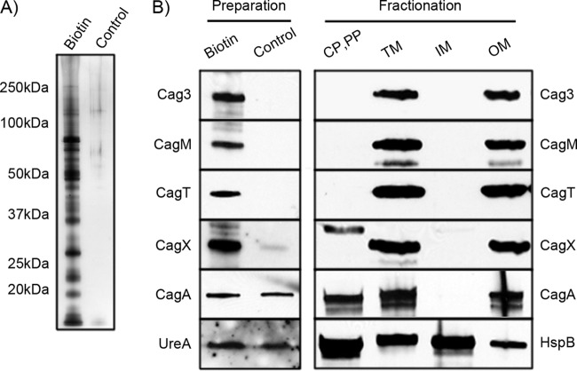
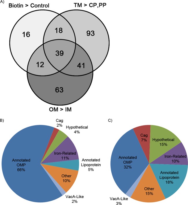
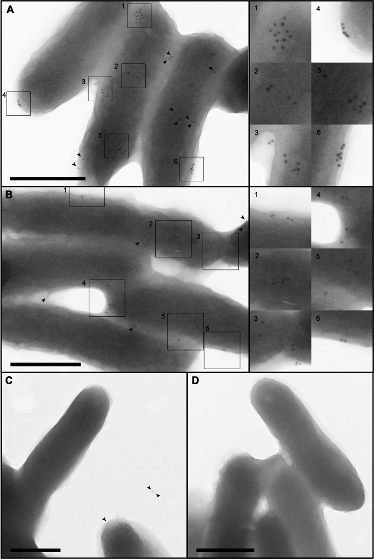
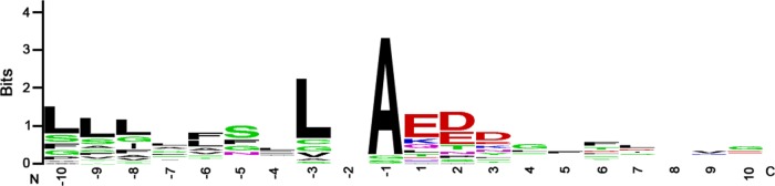
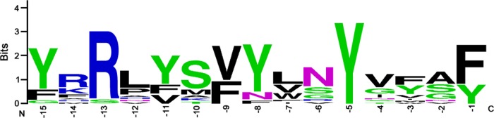
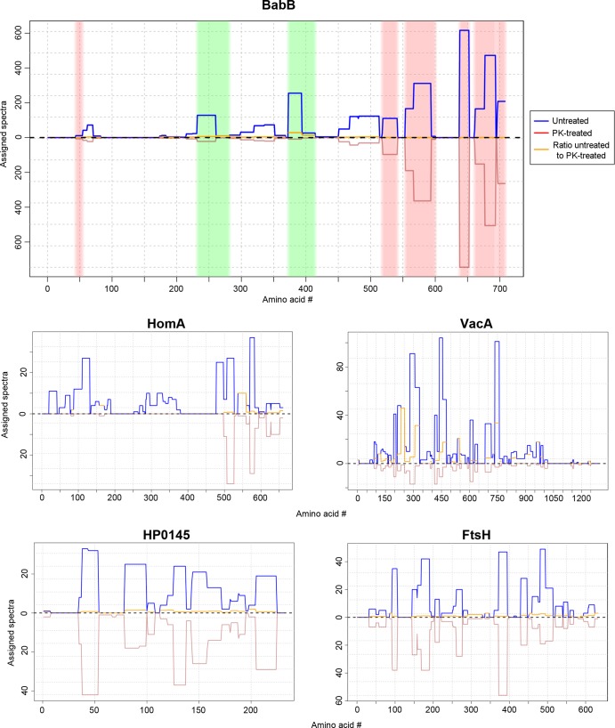
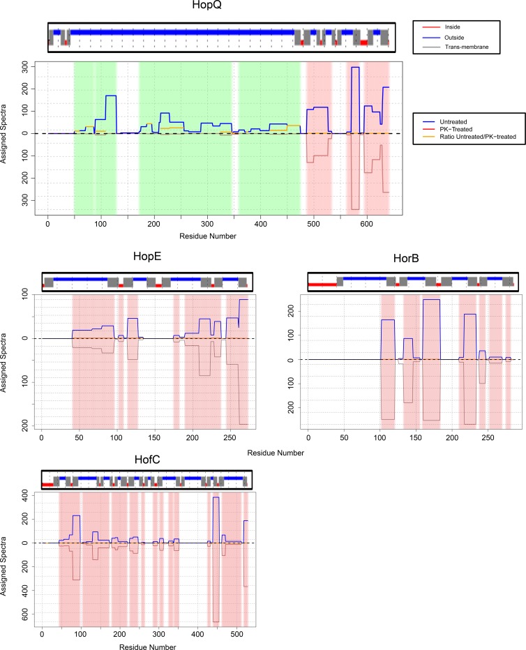
More than 50 Helicobacter pylori genes are predicted to encode outer membrane proteins (OMPs), but there has been relatively little experimental investigation of the H. pylori cell surface proteome. In this study, we used selective biotinylation to label proteins localized to the surface of H. pylori, along with differential detergent extraction procedures to isolate proteins localized to the outer membrane. Proteins that met multiple criteria for surface-exposed outer membrane localization included known adhesins, as well as Cag proteins required for activity of the cag type IV secretion system, putative lipoproteins, and other proteins not previously recognized as cell surface components. We identified sites of nontryptic cleavage consistent with signal sequence cleavage, as well as C-terminal motifs that may be important for protein localization. A subset of surface-exposed proteins were highly susceptible to proteolysis when intact bacteria were treated with proteinase K. Most Hop and Hom OMPs were susceptible to proteolysis, whereas Hor and Hof proteins were relatively resistant. Most of the protease-susceptible OMPs contain a large protease-susceptible extracellular domain exported beyond the outer membrane and a protease-resistant domain at the C terminus with a predicted β-barrel structure. These features suggest that, similar to the secretion of the VacA passenger domain, the N-terminal domains of protease-susceptible OMPs are exported through an autotransporter pathway. Collectively, these results provide new insights into the repertoire of surface-exposed H. pylori proteins that may mediate bacterium-host interactions, as well as the cell surface topology of these proteins.


Figures

FIG 1
FIG 1
Analysis of purified biotinylated proteins. (A) Biotinylated proteins were purified from biotinylated bacteria, and a control preparation was generated from nonbiotinylated bacteria. Equal volumes of each preparation were analyzed by silver staining. Specific bands were visible in the preparation of biotinylated proteins but not in the control preparation. The positions of molecular mass markers (in kilodaltons) are indicated to the left of the gel. (B) The preparations from panel A, as well as subcellular fractions, were immunoblotted with antisera to five proteins encoded by the cag PAI, UreA, or HspB. Equal volumes of biotinylated and unlabeled control preparations were loaded into each lane, and standardized amounts of subcellular fractions (25 μg of total protein) were loaded into each lane. The lanes contain soluble proteins predicted to have a cytoplasmic or periplasmic localization (CP,PP), insoluble proteins corresponding to a total membrane preparation (TM), Triton X-100-soluble membrane proteins predicted to have an inner membrane localization (IM), and Triton X-100-insoluble membrane proteins predicted to have an outer membrane localization (OM).
FIG 2
FIG 2
Use of multiple criteria to identify surface-exposed outer membrane proteins. (A) Venn diagram of proteins identified as enriched in the biotinylated preparation compared to the control preparation (Biotin > Control), enriched in the total membrane preparation compared to the soluble fraction (TM > CP,PP), and enriched in a Triton X-100-insoluble preparation compared to a Triton X-100-soluble preparation (OM > IM). Thirty-nine proteins met all three criteria. (B and C) Characteristics of the 39 proteins in the center segment of the Venn diagram, based on proportion of spectral counts (B) or proportion of annotated proteins in each class (C). Annotated OMPs represent Hop and Hor OMPs.
FIG 3
FIG 3
Localization of CagT by immunogold EM analysis. (A to C) H. pylori strains were immunolabeled with rabbit antiserum to CagT, followed by secondary antibodies conjugated to 10-nm gold particles. (A and B) Wild-type (WT) strain. (C) Δcag PAI mutant strain. Higher-magnification images of the regions in boxes, containing a high density of gold particles, are shown to the right of panels A and B. Arrowheads designate additional gold particles. (D) Immunogold labeling of the WT strain using secondary antibodies conjugated to gold particles, without primary antiserum. The number of gold particles detected on WT bacteria was significantly higher than the number detected on the cag ΔPAI mutant strain (mean 3.6 gold particles per WT cell and 1.8 gold particles per cag ΔPAI mutant cell, based on analysis of >200 bacteria of each type; P value < 0.0001, Welch two-sample t test). Bars, 500 nm.
FIG 4
FIG 4
Experimentally detected sites of signal peptide cleavage. We identified peptides in the preparations of biotinylated proteins that corresponded to non-tryptic-cleavage events and that could be matched to sites near the N termini of proteins, suggesting the occurrence of signal peptide cleavage. This analysis was restricted to peptides that could be matched to a single protein. Amino-terminal nontryptic cleavage was detected for 14 of the 39 surface-exposed proteins shown in Table 2 (see Table S6 in the supplemental material). These sequences were then aligned using WebLogo (weblogo.berkeley.edu). In the alignment, the numbering of positions is relative to the detected non-tryptic-cleavage site. The N terminus (N) and C terminus (C) are indicated.
FIG 5
FIG 5
Analysis of a conserved C-terminal motif. Among the 39 surface-exposed proteins shown in Table 2, 15 contain a distinctive C-terminal motif, characterized by conserved residues at positions −15, −13, −9, −5, and −1 (see Table S7 in the supplemental material). The numbers correspond to the distance from the carboxyl terminus. Sequences were aligned using WebLogo (weblogo.berkeley.edu).
FIG 6
FIG 6
Susceptibility of surface-exposed proteins to proteinase K treatment. Intact H. pylori cells were treated with proteinase K (1 mg ml−1) or buffer control at 4°C and then analyzed by MudPIT. Three experiments were performed, and the results were merged. We then analyzed the assigned spectra for each detected protein at the level of individual amino acids, and sites susceptible to proteolytic cleavage or resistant to proteolytic cleavage were identified, based on criteria defined in Materials and Methods. The blue lines (top half of graphs) depict the numbers of assigned spectra from experiments with control (untreated) bacteria, the red lines (bottom half of graphs) depict the numbers of assigned spectra from experiments with proteinase K (PK)-treated bacteria, and the orange lines indicate the ratio of assigned spectra in untreated compared to protease-treated bacteria. Data for 5 representative proteins are shown. In the top panel, the green shading illustrates protease-susceptible regions in BabB (exhibiting a ≥5-fold difference in the spectral counts when comparing untreated bacteria and protease-treated bacteria), and the pink shading illustrates protease-resistant regions (exhibiting a <2-fold difference in the spectral counts). The designation of protease-susceptible or protease-resistant regions was restricted to residues for which ≥5 assigned spectra were detected in the untreated samples. The N-terminal portions of BabB, HomA, and VacA were highly susceptible to protease digestion, whereas intracellular proteins such as the cytochrome c oxidase subunit FixO (HP0145) and the cell division protein FtsH were resistant to protease digestion.
FIG 7
FIG 7
Resistance of predicted β-barrel regions to digestion by proteinase K. This figure shows an analysis of the protease susceptibility of 4 H. pylori OMPs, using the approach shown in Fig. 6. The blue lines (top half of graphs) depict the numbers of assigned spectra from experiments with control (untreated) bacteria, and the red lines (bottom half of graphs) depict the numbers of assigned spectra from experiments with proteinase K-treated bacteria. The green shading illustrates protease-susceptible regions (exhibiting a ≥5-fold difference in the spectral counts when comparing untreated bacteria and protease-treated bacteria), and the pink shading illustrates protease-resistant regions (exhibiting a <2-fold difference in the spectral counts). The designation of protease-susceptible or protease-resistant regions was restricted to residues for which ≥5 assigned spectra were detected in the untreated samples. The locations of transmembrane β-strands were predicted using the program BOCTOPUS and are shown above each graph (red indicates predicted periplasm-facing residues, gray indicates predicted transmembrane β-strands, and blue indicates predicted extracellular facing residues). A large N-terminal portion of HopQ is highly susceptible to proteolytic digestion, whereas the C-terminal portion of HopQ is resistant to proteolytic digestion. The other 3 OMPs (HopE, HorB, and HofC) are relatively resistant to proteolysis. The resistant C-terminal region of HopQ corresponds to a region predicted by BOCTOPUS to have multiple transmembrane β-strands, consistent with a β-barrel structure. Three protease-resistant OMPs (HopE, HorB, and HofC) are each predicted to have predominantly β-barrel structure.

References

    1. Atherton JC, Blaser MJ. 2009. Coadaptation of Helicobacter pylori and humans: ancient history, modern implications. J. Clin. Invest. 119:2475–2487. 10.1172/JCI38605 - DOI - PMC - PubMed
    1. Cover TL, Blaser MJ. 2009. Helicobacter pylori in health and disease. Gastroenterology 136:1863–1873. 10.1053/j.gastro.2009.01.073 - DOI - PMC - PubMed
    1. Amieva MR, El-Omar EM. 2008. Host-bacterial interactions in Helicobacter pylori infection. Gastroenterology 134:306–323. 10.1053/j.gastro.2007.11.009 - DOI - PubMed
    1. Polk DB, Peek RM., Jr 2010. Helicobacter pylori: gastric cancer and beyond. Nat. Rev. Cancer 10:403–414. 10.1038/nrc2857 - DOI - PMC - PubMed
    1. Cover TL, Blanke SR. 2005. Helicobacter pylori VacA, a paradigm for toxin multifunctionality. Nat. Rev. Microbiol. 3:320–332. 10.1038/nrmicro1095 - DOI - PubMed

Publication types

MeSH terms

Substances