. 2014 Aug;21(8):1250-61.
doi: 10.1038/cdd.2014.44. Epub 2014 Apr 25.

Identification of a ZEB2-MITF-ZEB1 transcriptional network that controls melanogenesis and melanoma progression

Affiliations

Identification of a ZEB2-MITF-ZEB1 transcriptional network that controls melanogenesis and melanoma progression

G Denecker et al. Cell Death Differ. 2014 Aug.

Abstract

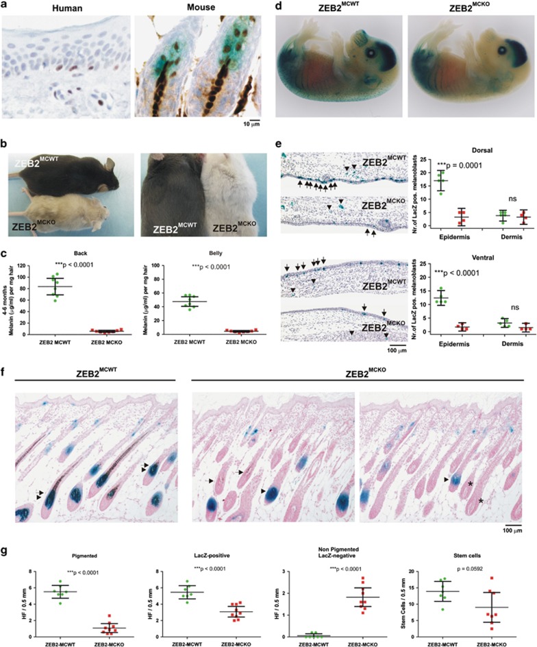
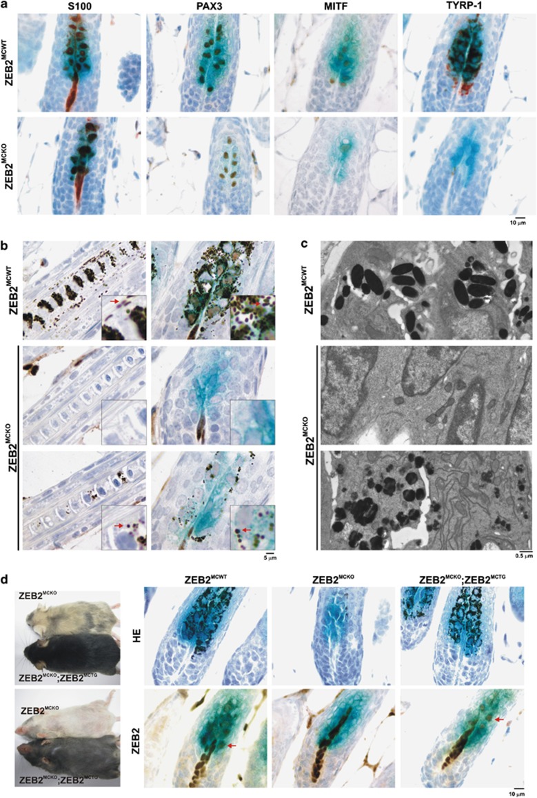
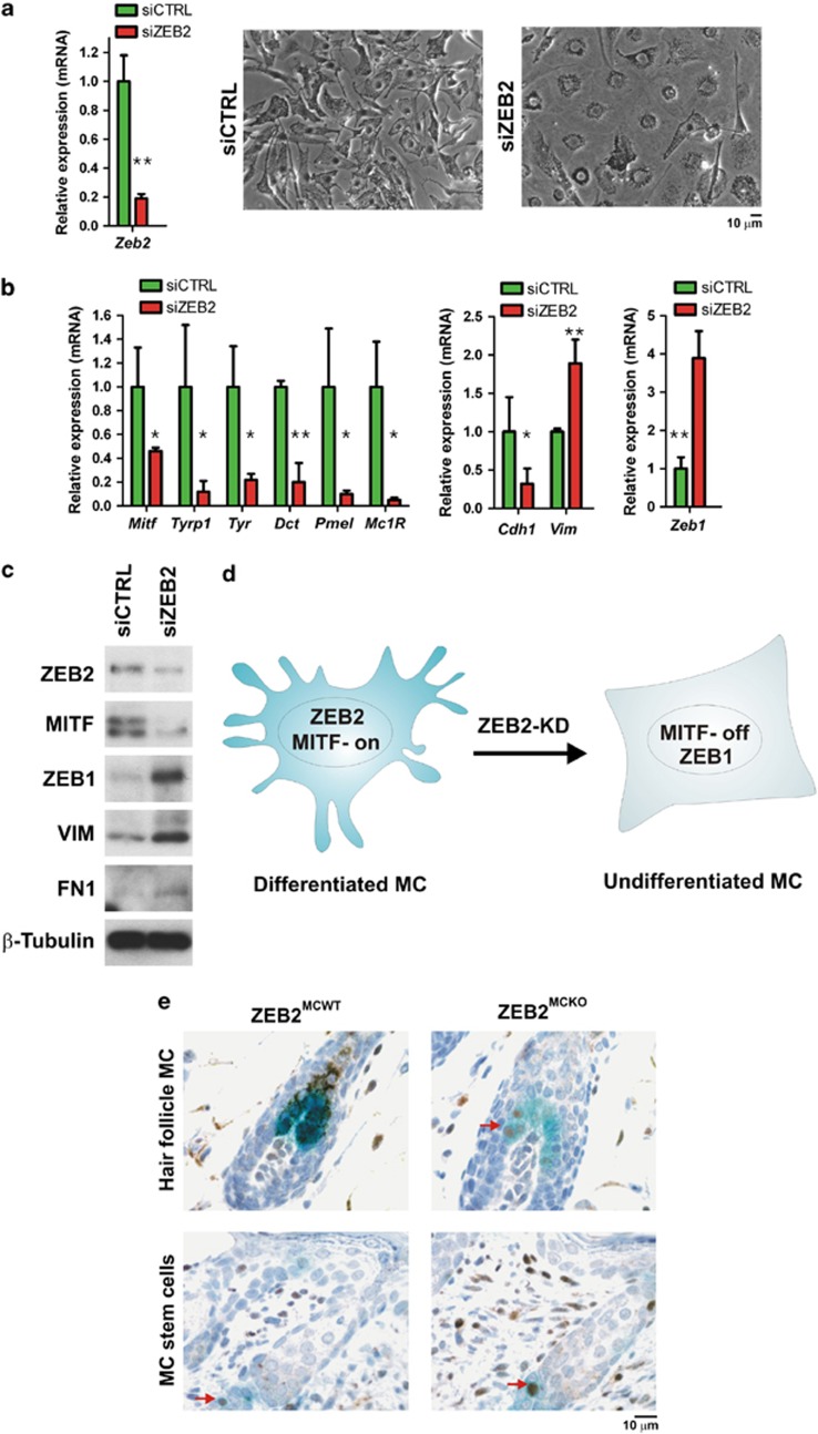
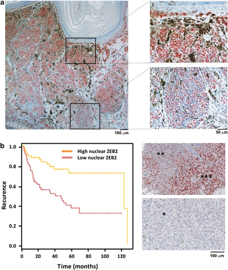
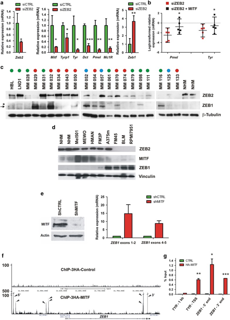
Deregulation of signaling pathways that control differentiation, expansion and migration of neural crest-derived melanoblasts during normal development contributes also to melanoma progression and metastasis. Although several epithelial-to-mesenchymal (EMT) transcription factors, such as zinc finger E-box binding protein 1 (ZEB1) and ZEB2, have been implicated in neural crest cell biology, little is known about their role in melanocyte homeostasis and melanoma. Here we show that mice lacking Zeb2 in the melanocyte lineage exhibit a melanoblast migration defect and, unexpectedly, a severe melanocyte differentiation defect. Loss of Zeb2 in the melanocyte lineage results in a downregulation of the Microphthalmia-associated transcription factor (Mitf) and melanocyte differentiation markers concomitant with an upregulation of Zeb1. We identify a transcriptional signaling network in which the EMT transcription factor ZEB2 regulates MITF levels to control melanocyte differentiation. Moreover, our data are also relevant for human melanomagenesis as loss of ZEB2 expression is associated with reduced patient survival.


Figures

Figure 1
Figure 1
Zeb2 loss in the melanocyte lineage causes congenital loss of pigmentation and impairs melanoblast development. (a) ZEB2 is expressed in differentiated melanocytes in the human epidermis, and in the hair follicles of mouse skin. (b) Melanocyte-specific deletion of Zeb2 (ZEB2MCKO) causes congenital loss of hair pigmentation in homozygous knockout mice (2 months old). This loss of pigmentation was more pronounced on the belly than on the back. (c) Determination of melanin pigment (μg/ml) in the dorsal and ventral hair of ZEB2MCWT and ZEB2MCKO mice (n=7–8 for each group) at the age of 4–6 months. (d) ZEB2 is required for proper melanoblast development in homozygous embryos at E15.5. Whole-mount LacZ staining of E15.5 ZEB2MCWT;Dct-LacZ and ZEB2MCKO;Dct-LacZ embryos. (e) Comparison of melanoblast migration in the epidermis and dermis of dorsal and ventral areas of ZEB2MCWT;Dct-LacZ and ZEB2MCKO;Dct-LacZ embryos (n=4 and 5, respectively). Results represent the number of melanoblasts per area analyzed. (f) Nuclear fast-red staining of LacZ-stained skin sections of ZEB2MCWT;Dct-LacZ and ZEB2MCKO;Dct-LacZ mice to visualize hair follicles (HF) (arrow: pigmented HF, arrowhead: LacZ-positive HF, *: LacZ-negative HF). (g) Quantitative analysis of the pigmented, LacZ-positive, non-pigmented LacZ-negative HF and LacZ-positive stem cells in the bulge area of ZEB2MCWT (n=7) and ZEB2MCKO (n=8) mice per 0.5 mm analyzed. Data of ZEB2MCWT and ZEB2MCKOmice were compared by using unpaired Student's t-test and are presented as means±95% CI. P-values are indicated with (P<0.001). Micrograph images were taken with a × 60/0.8 objective (a) or a × 10/0.25 objective (e and f)
Figure 2
Figure 2
Melanocyte-specific Zeb2 deficiency causes the formation of undifferentiated melanocytes in the bulge area of hair follicles. (a) Immunohistochemical staining of sections of ZEB2MCWT and ZEB2MCKO;Dct-LacZ-positive skin sections with S100b, PAX3, MITF and TYRP1 antibodies. (b) Detailed microscopic analysis of melanosomes in the hair shafts and the bulb area of the hair follicles, combined with immunohistochemical staining of ZEB2 on LacZ-positive skin sections of ZEB2MCWT and ZEB2MCKO; Dct-LacZ-positive mice. Insets show the altered morphology of melanosomes in the ZEB2MCKO sections compared with ZEB2MCWT. (c) Electron microscopic analysis of melanosomes in the bulb area of ZEB2MCWT and ZEB2MCKO mice demonstrates the absence or irregular morphology of melanosomes in the ZEB2MCKO hair follicles. (d) Genetic compensation of the loss of Zeb2 in the ZEB2MCKO;Dct-LacZ mice with melanocyte-specific overexpression of ZEB2 (ZEB2MCTG). Left panel: complete compensation of pigmentation in ZEB2MCKO ZEB2MCTG;Dct-LacZ mice compared with ZEB2MCKO;Dct-LacZ mice. Right panels: reappearance of pigmented melanosomes and ZEB2 expression in the ZEB2MCKO ZEB2MCTG;Dct-LacZ hair follicles. All microscopic analyses were done on skin sections of 5.5-day-old (ac) or 13.5-day-old mice (d) and Immunohistochemical micrograph images were taken with a × 60/0.8 objective (a and d) or a × 100/1.25 objective (b)
Figure 3
Figure 3
ZEB2 is necessary for proper differentiation of primary melanocytes. (a and b) Morphology of primary melanocytes after knockdown of Zeb2 by siRNA transfection and relative mRNA expression of Zeb2, melanocytes-specific differentiation genes Tyrp1, Tyr, Dct, PmeL and Mc1R, the epithelial/mesenchymal markers E-cadherin/Vimentin and Zeb1. Scrambled siRNA was used as a control (siCTRL). (c) Western blot analysis after siRNA knockdown of Zeb2 in the primary Melan a cell line. (d) Hypothetical model of the ZEB2-MITF-ZEB1 balance in melanocytes (e) ZEB1 expression in dedifferentiated LacZ-positive melanocytes of the hair follicles of ZEB2MCKO mice and in the melanocyte stem cells of both ZEB2MCWT and ZEB2MCKO mice. QPCR data were compared by using unpaired Student's t-test (n=3) and are presented as averages±S.D. P-values are indicated with (**P<0.005 and *P<0.05). Micrograph images were taken with a × 20/0.2 objective (a) or a × 60/0.8 objective (e)
Figure 4
Figure 4
ZEB2 expression in melanoma. (a) Heterogeneous ZEB2 expression in primary human melanoma with vertical growth phase. (b) Kaplan–Meier curves for melanoma recurrence-free survival, comparing human melanoma samples with strong nuclear ZEB2 expression (n=83) and weak nuclear ZEB2 expression (n=82) from a melanoma tissue array. Data were compared using the log-rank test and **P=0.0065. A representative ZEB2 staining of both groups is shown in the right panel (*** very high nuclear ZEB2, ** high nuclear ZEB2, * low nuclear ZEB2). ZEB2 protein expression values were determined using an automated image analysis approach (IHC-MARK, Oncomark, Dublin, Ireland) designed to quantify immunohistochemically stained slides. Micrograph images were taken with a × 4/0.1 and × 20/0.4 objective (a) or a × 10/0.25 objective (b)
Figure 5
Figure 5
The ZEB2-MITF-ZEB1 transcriptional network. (a) Knockdown of Zeb2 by siRNA transfection of the B16 melanoma cell line. Scrambled siRNA was used as a control (siCTRL). Relative mRNA expression of endogenous Zeb2, the melanocyte-specific differentiation genes Mitf, Tyrp1, Tyr, Dct, Mc1R and Zeb1 after Zeb2 knockdown are shown. (b) Relative mRNA expression of subset of MITF target genes after siRNA knockdown of Zeb2 in the B16 melanoma cell line after compensation with Mitf. (c) Western blot analysis of ZEB2 and ZEB1 expression in short-term culture human melanoma cell lines with different NRAS (blue circles) or BRAF (red circles) mutated or WT (green circles) status. Primary human melanocytes were used as a reference. (→) specific ZEB1 band, (*) aspecific band. (d) Western blot analysis of ZEB2, MITF and ZEB1 in a panel of human melanoma cell lines. Primary human melanocytes were used as a reference. (e) 501Mel cells were infected with lentiviral vectors expressing control shRNA (shCTRL) or shRNA directed against MITF (shMITF). Left panel: western blot analysis shows strongly downregulated MITF expression after shMITF infection. Right panel: shMITF knockdown leads to increased ZEB1 mRNA expression. Two independent primer pairs located in exons 1–2 and 4–5 for the ZEB1 gene were used. (f) UCSC screenshot of a wig file of an HA-ChIP-seq from 501Mel cells expressing a 3HA-Control vector or 3HA-tagged MITF at the ZEB1 locus. Several MITF binding sites are observed (shown by arrows) both upstream and downstream of the ZEB1 gene. (g) Anti-HA-ChIP-qPCR from native 501Mel cells (CTRL) or 501MEL cells stably expressing 3HA-tagged MITF (HA-MITF). qPCR was performed with primers that amplify a region 1 kb upstream of the TYR transcription start site (TSS) as negative control, the TYR TSS as positive control and two sites from the ZEB1 locus 5′ and 3′ of the gene as indicated in f. QPCR data were compared by using unpaired Student's t-test (n=3, for a, e and g) or paired Student's t-test (n=5, for b) and are presented as averages±S.D. or are presented as log-transformed relative expression values with 95% CI. P-values are indicated with ***P<0.0005, **P<0.005 and *P<0.05

Similar articles

Cited by

References

    1. Ernfors P. Cellular origin and developmental mechanisms during the formation of skin melanocytes. Exp Cell Res. 2010;316:1397–1407. - PubMed
    1. Luciani F, Champeval D, Herbette A, Denat L, Aylaj B, Martinozzi S, et al. Biological and mathematical modeling of melanocyte development. Development. 2011;138:3943–3954. - PubMed
    1. Osawa M.Melanocyte stem cells(June 30, 2009), StemBook, ed. The Stem Cell Research Community, StemBook,doi:10.3824/stembook.1.46.1 - DOI
    1. Pshenichnaya I, Schouwey K, Armaro M, Larue L, Knoepfler PS, Eisenman RN, et al. Constitutive gray hair in mice induced by melanocyte-specific deletion of c-Myc. Pigment Cell Melanoma Res. 2012;25:312–325. - PMC - PubMed
    1. Steingrimsson E, Copeland NG, Jenkins NA. Melanocyte stem cell maintenance and hair graying. Cell. 2005;121:9–12. - PubMed

Publication types

MeSH terms

Substances