Complex proteinopathy with accumulations of prion protein, hyperphosphorylated tau, α-synuclein and ubiquitin in experimental bovine spongiform encephalopathy of monkeys
- PMID: 24769839
- PMCID: PMC4059271
- DOI: 10.1099/vir.0.062083-0
Complex proteinopathy with accumulations of prion protein, hyperphosphorylated tau, α-synuclein and ubiquitin in experimental bovine spongiform encephalopathy of monkeys
Abstract
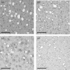
Proteins aggregate in several slowly progressive neurodegenerative diseases called 'proteinopathies'. Studies with cell cultures and transgenic mice overexpressing mutated proteins suggested that aggregates of one protein induced misfolding and aggregation of other proteins as well - a possible common mechanism for some neurodegenerative diseases. However, most proteinopathies are 'sporadic', without gene mutation or overexpression. Thus, proteinopathies in WT animals genetically close to humans might be informative. Squirrel monkeys infected with the classical bovine spongiform encephalopathy agent developed an encephalopathy resembling variant Creutzfeldt-Jakob disease with accumulations not only of abnormal prion protein (PrP(TSE)), but also three other proteins: hyperphosphorylated tau (p-tau), α-synuclein and ubiquitin; β-amyloid protein (Aβ) did not accumulate. Severity of brain lesions correlated with spongiform degeneration. No amyloid was detected. These results suggested that PrP(TSE) enhanced formation of p-tau and aggregation of α-synuclein and ubiquitin, but not Aβ, providing a new experimental model for neurodegenerative diseases associated with complex proteinopathies.
Figures


Similar articles
-
Experimental Bovine Spongiform Encephalopathy in Squirrel Monkeys: The Same Complex Proteinopathy Appearing after Very Different Incubation Times.Pathogens. 2022 May 20;11(5):597. doi: 10.3390/pathogens11050597. Pathogens. 2022. PMID: 35631118 Free PMC article.
-
Squirrel monkeys (Saimiri sciureus) infected with the agent of bovine spongiform encephalopathy develop tau pathology.J Comp Pathol. 2012 Jul;147(1):84-93. doi: 10.1016/j.jcpa.2011.09.004. Epub 2011 Oct 20. J Comp Pathol. 2012. PMID: 22018806 Free PMC article.
-
Identification of a second bovine amyloidotic spongiform encephalopathy: molecular similarities with sporadic Creutzfeldt-Jakob disease.Proc Natl Acad Sci U S A. 2004 Mar 2;101(9):3065-70. doi: 10.1073/pnas.0305777101. Epub 2004 Feb 17. Proc Natl Acad Sci U S A. 2004. PMID: 14970340 Free PMC article.
-
Insights into Mechanisms of Transmission and Pathogenesis from Transgenic Mouse Models of Prion Diseases.Methods Mol Biol. 2017;1658:219-252. doi: 10.1007/978-1-4939-7244-9_16. Methods Mol Biol. 2017. PMID: 28861793 Free PMC article. Review.
-
How an Infection of Sheep Revealed Prion Mechanisms in Alzheimer's Disease and Other Neurodegenerative Disorders.Int J Mol Sci. 2021 May 4;22(9):4861. doi: 10.3390/ijms22094861. Int J Mol Sci. 2021. PMID: 34064393 Free PMC article. Review.
Cited by
-
Aggregation of MBP in chronic demyelination.Ann Clin Transl Neurol. 2015 Jul;2(7):711-21. doi: 10.1002/acn3.207. Epub 2015 Jun 6. Ann Clin Transl Neurol. 2015. PMID: 26273684 Free PMC article.
-
Experimental Bovine Spongiform Encephalopathy in Squirrel Monkeys: The Same Complex Proteinopathy Appearing after Very Different Incubation Times.Pathogens. 2022 May 20;11(5):597. doi: 10.3390/pathogens11050597. Pathogens. 2022. PMID: 35631118 Free PMC article.
-
Exploration of the Main Sites for the Transformation of Normal Prion Protein (PrPC) into Pathogenic Prion Protein (PrPsc).J Vet Res. 2017 Apr 4;61(1):11-22. doi: 10.1515/jvetres-2017-0002. eCollection 2017 Mar. J Vet Res. 2017. PMID: 29978050 Free PMC article.
-
A Naturally Occurring Bovine Tauopathy Is Geographically Widespread in the UK.PLoS One. 2015 Jun 19;10(6):e0129499. doi: 10.1371/journal.pone.0129499. eCollection 2015. PLoS One. 2015. PMID: 26091261 Free PMC article.
References
Publication types
MeSH terms
Substances
Grants and funding
LinkOut - more resources
Full Text Sources
Other Literature Sources
Research Materials

