

Stacked stem cell sheets enhance cell-matrix interactions
- PMID: 24769850
- PMCID: PMC4154950
- DOI: 10.4161/org.28990
Stacked stem cell sheets enhance cell-matrix interactions
Abstract
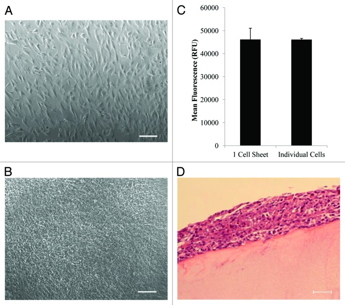
Cell sheet engineering has enabled the production of confluent cell sheets stacked together for use as a cardiac patch to increase cell survival rate and engraftment after transplantation, thereby providing a promising strategy for high density stem cell delivery for cardiac repair. One key challenge in using cell sheet technology is the difficulty of cell sheet handling due to its weak mechanical properties. A single-layer cell sheet is generally very fragile and tends to break or clump during harvest. Effective transfer and stacking methods are needed to move cell sheet technology into widespread clinical applications. In this study, we developed a simple and effective micropipette based method to aid cell sheet transfer and stacking. The cell viability after transfer was tested and multi-layer stem cell sheets were fabricated using the developed method. Furthermore, we examined the interactions between stacked stem cell sheets and fibrin matrix. Our results have shown that the preserved ECM associated with the detached cell sheet greatly facilitates its adherence to fibrin matrix and enhances the cell sheet-matrix interactions. Accelerated fibrin degradation caused by attached cell sheets was also observed.
Keywords: cell sheet; cell-matrix interactions; fibrin; mesenchymal stem cells.
Figures


Similar articles
-
Effective stacking and transplantation of stem cell sheets using exogenous ROS-producing film for accelerated wound healing.Acta Biomater. 2019 Sep 1;95:418-426. doi: 10.1016/j.actbio.2019.01.019. Epub 2019 Jan 16. Acta Biomater. 2019. PMID: 30660002
-
Phenotypic traits of mesenchymal stem cell sheets fabricated by temperature-responsive cell culture plate: structural characteristics of MSC sheets.Stem Cell Res Ther. 2019 Nov 28;10(1):353. doi: 10.1186/s13287-019-1431-6. Stem Cell Res Ther. 2019. PMID: 31779694 Free PMC article.
-
Reliable Harvest of Injectable Human Mesenchymal Stem Cell Sheets by Modulating Cell-Substrate Adhesion Strength.Adv Healthc Mater. 2025 May;14(14):e2500135. doi: 10.1002/adhm.202500135. Epub 2025 Apr 18. Adv Healthc Mater. 2025. PMID: 40249130
-
The preparation methods and types of cell sheets engineering.Stem Cell Res Ther. 2024 Sep 27;15(1):326. doi: 10.1186/s13287-024-03937-4. Stem Cell Res Ther. 2024. PMID: 39334404 Free PMC article. Review.
-
Mesenchymal stem cell sheets: a new cell-based strategy for bone repair and regeneration.Biotechnol Lett. 2019 Mar;41(3):305-318. doi: 10.1007/s10529-019-02649-7. Epub 2019 Jan 24. Biotechnol Lett. 2019. PMID: 30680496 Review.
Cited by
-
Immunobiology of fibrin-based engineered heart tissue.Stem Cells Transl Med. 2015 Jun;4(6):625-31. doi: 10.5966/sctm.2013-0202. Epub 2015 May 6. Stem Cells Transl Med. 2015. PMID: 25947338 Free PMC article.
-
Tailoring cell sheets for biomedical applications.Smart Med. 2024 Feb 18;3(1):e20230038. doi: 10.1002/SMMD.20230038. eCollection 2024 Feb. Smart Med. 2024. PMID: 39188516 Free PMC article. Review.
-
Enhancing chondrogenic potential via mesenchymal stem cell sheet multilayering.Regen Ther. 2021 Dec 2;18:487-496. doi: 10.1016/j.reth.2021.11.004. eCollection 2021 Dec. Regen Ther. 2021. PMID: 34926734 Free PMC article.
-
UTP Regulates the Cardioprotective Action of Transplanted Stem Cells Derived From Mouse Cardiac Adipose Tissue.Front Pharmacol. 2022 Jun 15;13:906173. doi: 10.3389/fphar.2022.906173. eCollection 2022. Front Pharmacol. 2022. PMID: 35784739 Free PMC article.
-
Scaffold-free cell-based tissue engineering therapies: advances, shortfalls and forecast.NPJ Regen Med. 2021 Mar 29;6(1):18. doi: 10.1038/s41536-021-00133-3. NPJ Regen Med. 2021. PMID: 33782415 Free PMC article. Review.
References
-
- Roger VL, Go AS, Lloyd-Jones DM, Benjamin EJ, Berry JD, Borden WB, Bravata DM, Dai S, Ford ES, Fox CS, et al. American Heart Association Statistics Committee and Stroke Statistics Subcommittee Executive summary: heart disease and stroke statistics--2012 update: a report from the American Heart Association. Circulation. 2012;125:188–97. doi: 10.1161/CIR.0b013e3182456d46. - DOI - PubMed
-
- Caulfield JB, Leinbach R, Gold H. The relationship of myocardial infarct size and prognosis. Circulation. 1976;53(Suppl):I141–4. - PubMed
Publication types
MeSH terms
Substances
Grants and funding
LinkOut - more resources
Full Text Sources
Other Literature Sources