

The special case of hepatocytes: unique tissue architecture calls for a distinct mode of cell division
- PMID: 24769852
- PMCID: PMC4199811
- DOI: 10.4161/bioa.29012
The special case of hepatocytes: unique tissue architecture calls for a distinct mode of cell division
Abstract
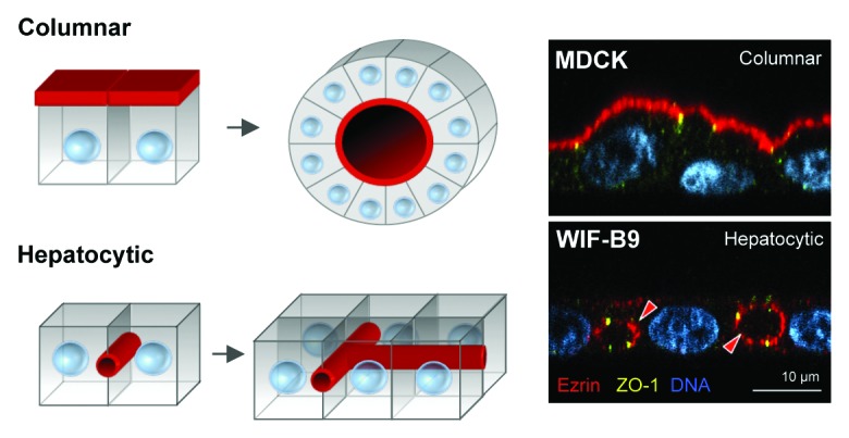
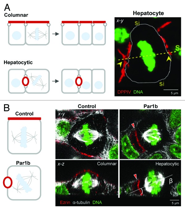
Columnar epithelia (e.g., kidney, intestine) and hepatocytes embody the two major organizational phenotypes of non-stratified epithelial cells. Columnar epithelia establish their apical and basal domains at opposing poles and organize in monolayered cysts and tubules, in which their apical surfaces form a single continuous lumen whereas hepatocytes establish their apical domains in the midst of their basolateral domains and organize a highly branched capillary luminal network, the bile canaliculi, in which a single hepatocyte can engage in lumen formation with multiple neighbors. To maintain their distinct tissue architectures, columnar epithelial cells bisect their luminal domains during symmetric cell divisions, while the cleavage furrow in dividing hepatocytes avoids bisecting the bile canalicular domains. We discuss recently discovered molecular mechanisms that underlie the different cell division phenotypes in columnar and hepatocytic model cell lines. The serine/threonine kinase Par1b determines both the epithelial lumen polarity and cell division phenotype via cell adhesion signaling that converges on the small GTPase RhoA.
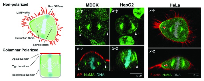
Keywords: LGN/NuMA; Par1b/MARK2; RhoA; columnar and hepatocyte polarity; epithelial cells; hepatocyte polarity; mitotic spindle orientation.
Figures
Similar articles
-
Par1b links lumen polarity with LGN-NuMA positioning for distinct epithelial cell division phenotypes.J Cell Biol. 2013 Oct 28;203(2):251-64. doi: 10.1083/jcb.201303013. J Cell Biol. 2013. PMID: 24165937 Free PMC article.
-
Par1b promotes hepatic-type lumen polarity in Madin Darby canine kidney cells via myosin II- and E-cadherin-dependent signaling.Mol Biol Cell. 2007 Jun;18(6):2203-15. doi: 10.1091/mbc.e07-02-0095. Epub 2007 Apr 4. Mol Biol Cell. 2007. PMID: 17409351 Free PMC article.
-
Cell-cell adhesion accounts for the different orientation of columnar and hepatocytic cell divisions.J Cell Biol. 2017 Nov 6;216(11):3847-3859. doi: 10.1083/jcb.201608065. Epub 2017 Sep 8. J Cell Biol. 2017. PMID: 28887437 Free PMC article.
-
Hepatocyte polarity.Compr Physiol. 2013 Jan;3(1):243-87. doi: 10.1002/cphy.c120009. Compr Physiol. 2013. PMID: 23720287 Free PMC article. Review.
-
The unique polarity phenotype of hepatocytes.Exp Cell Res. 2014 Nov 1;328(2):276-83. doi: 10.1016/j.yexcr.2014.06.006. Epub 2014 Jun 20. Exp Cell Res. 2014. PMID: 24956563 Free PMC article. Review.
Cited by
-
Impact of HCV Infection on Hepatocyte Polarity and Plasticity.Pathogens. 2022 Mar 10;11(3):337. doi: 10.3390/pathogens11030337. Pathogens. 2022. PMID: 35335661 Free PMC article. Review.
-
Induction of Bile Canaliculi-Forming Hepatocytes from Human Pluripotent Stem Cells.Methods Mol Biol. 2022;2544:71-82. doi: 10.1007/978-1-0716-2557-6_4. Methods Mol Biol. 2022. PMID: 36125710
-
Optimization of extracellular matrix for primary human hepatocyte cultures using mixed collagen-Matrigel matrices.EXCLI J. 2023 Jan 4;22:12-34. doi: 10.17179/excli2022-5459. eCollection 2023. EXCLI J. 2023. PMID: 36660192 Free PMC article.
-
Ultrastructural Features of Gold Nanoparticles Interaction with HepG2 and HEK293 Cells in Monolayer and Spheroids.Nanomaterials (Basel). 2020 Oct 16;10(10):2040. doi: 10.3390/nano10102040. Nanomaterials (Basel). 2020. PMID: 33081137 Free PMC article.
-
"iPSC-derived liver organoids and inherited bleeding disorders: Potential and future perspectives".Front Physiol. 2023 Jan 13;14:1094249. doi: 10.3389/fphys.2023.1094249. eCollection 2023. Front Physiol. 2023. PMID: 36711019 Free PMC article. Review.
References
Publication types
MeSH terms
Substances
Grants and funding
LinkOut - more resources
Full Text Sources
Other Literature Sources
Research Materials
Miscellaneous