. 2014;13(12):1987-94.
doi: 10.4161/cc.28929. Epub 2014 Apr 25.

Coffee induces autophagy in vivo

Affiliations

Coffee induces autophagy in vivo

Federico Pietrocola et al. Cell Cycle. 2014.

Abstract

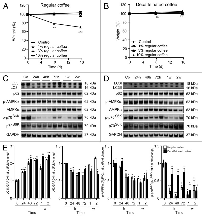
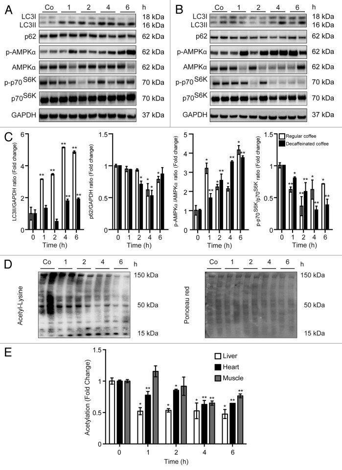
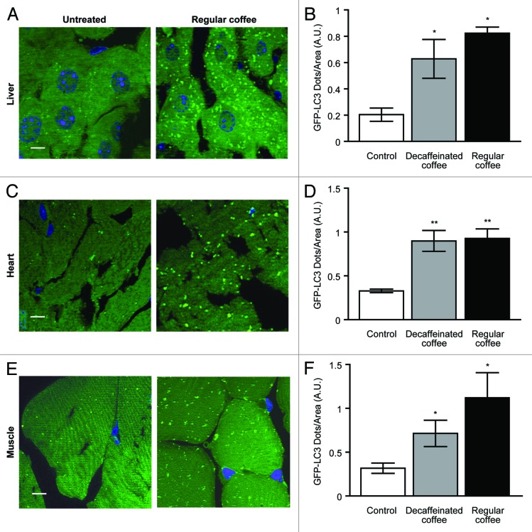
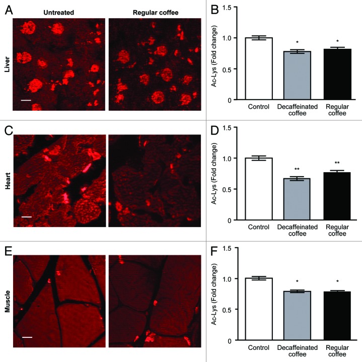
Epidemiological studies and clinical trials revealed that chronic consumption coffee is associated with the inhibition of several metabolic diseases as well as reduction in overall and cause-specific mortality. We show that both natural and decaffeinated brands of coffee similarly rapidly trigger autophagy in mice. One to 4 h after coffee consumption, we observed an increase in autophagic flux in all investigated organs (liver, muscle, heart) in vivo, as indicated by the increased lipidation of LC3B and the reduction of the abundance of the autophagic substrate sequestosome 1 (p62/SQSTM1). These changes were accompanied by the inhibition of the enzymatic activity of mammalian target of rapamycin complex 1 (mTORC1), leading to the reduced phosphorylation of p70(S6K), as well as by the global deacetylation of cellular proteins detectable by immunoblot. Immunohistochemical analyses of transgenic mice expressing a GFP-LC3B fusion protein confirmed the coffee-induced relocation of LC3B to autophagosomes, as well as general protein deacetylation. Altogether, these results indicate that coffee triggers 2 phenomena that are also induced by nutrient depletion, namely a reduction of protein acetylation coupled to an increase in autophagy. We speculate that polyphenols contained in coffee promote health by stimulating autophagy.

Keywords: acetyl-coenzyme A; acetylation; mTOR; macroautophagy.


Figures

None
Figure 1. Long-term administration of regular and decaffeinated coffee at a dose not affecting body weight induces autophagy in the liver. (A and B). Effect of 2 wk administration of regular (A) or decaffeinated coffee (B) on body weight. C57BL/6 mice were administrated with the indicated doses of regular coffee (A) or decaffeinated coffee (B) diluted in drinking water, which was provided to mice ad libitum. The dose not affecting body weight (3% w/v) was designated for investigating pro-autophagic effects. Results from n = 3 independent experiments. (C and D). Immunoblotting analysis of long-term coffee administration on autophagy regulation in liver. Administration of both regular (C) and decaffeinated coffee (D) for up to 2 wk (w) resulted in activation of autophagy, as measured by LC3 lipidation and p62/SQSTM1 degradation (quantified in E). Autophagy activation was associated with a decrease in mTORC1 activity, as measured by the phosphorylation of p70s6k, but not to an activation of AMPK (quantified in E). GAPDH levels were monitored to ensure equal loading. Representative images are reported in (CandD). Results from n = 3 independent experiments are presented as fold change ± SEM *P < 0.05; **P < 0.01; ***P < 0.005 (unpaired, 2-tailed Student t test), compared with untreated mice.
None
Figure 2. Short-term administration of both regular coffee and decaffeinated coffee induces autophagy accompanied by a reduction in global acetylation levels of proteins in the liver. (A and B). Immunoblotting analysis of short-term coffee administration on autophagy regulation in liver. Gavage of both regular coffee (A) and decaffeinated coffee (B) resulted in an activation of autophagy, although at different extent and timing, as measured by LC3 lipidation and p62 degradation (quantified in C). In both cases, autophagy induction was accompanied by an activation of AMPK and by a reduction in the activity of mTORC1, as measured by the phosphorylation of its substrate p70s6k (quantified in C). Representative images are depicted in (AandB). Results from n = 3 independent experiments are presented as fold change ± SEM *P < 0.05; **P < 0.01; (unpaired, 2-tailed Student t test), compared with untreated mice. (D and E) Immunoblot detection of protein acetylation in mice administered with regular coffee. Coffee administration (by gavage) resulted in a significant drop in the overall acetylation levels of proteins in liver, heart, and muscle (quantified in E) in a range of time of 1 to 6 h depending on the tissue. Panels in (D) refer to liver. Ponceau red staining was used to monitor equal loading of the lanes. Results from n = 3 independent experiments are presented as fold change ± SEM *P < 0.05; **P < 0.01 (unpaired, 2-tailed Student t test), compared with untreated mice.
None
Figure 3. Autophagy induction mediated by short-term coffee administration is confirmed for liver, heart, and muscle from GFP-LC3 transgenic mice. GFP-LC3-expressing mice were administered with regular and decaffeinated coffee by gavage and analyzed for autophagy induction after 4 h. (A–F). Fluorescence microscopic analysis of liver (A), heart (C), and muscle (E) revealed a significant increase in the number of LC3II puncta per area of cells in all tissues (quantified in B, D, andF). Representative images of tissues from untreated vs. regular coffee-treated mice are depicted in (A, C, and E) (bar scale: 10 µm). Results from 3 independent experiments are presented as GFP-LC3 dots/area (means ± SEM). *P < 0.05; **P < 0.01; (unpaired, 2-tailed Student t test), compared with untreated mice.
None
Figure 4. Reduction in global protein acetylation levels mediated by short-term coffee administration in liver, heart, and muscle. Livers, hearts, and muscles from C57Bl/6 mice were analyzed 4 h after treatment with regular and decaffeinated coffee to determine protein acetylatation by immunofluorescence. Fluorescence microscope analysis of liver (A), heart (C), and muscle (E) revealed a significant decrease in the acetylation of proteins in all tissues (quantified in B, D, andF). Representative images of untreated vs. regular coffee-treated mice are depicted in (A, C, and E) (bar scale: 10 µm). Results from n = 3 independent experiments are presented as acetylated lysine intensity fold change ± SEM, considering the values of tissues from untreated mice as 1. *P < 0.05; **P < 0.01; (unpaired, 2-tailed Student t test), compared with untreated mice.

Comment in

  • The cup of youth.
    Dumit VI, Dengjel J. Dumit VI, et al. Cell Cycle. 2014;13(13):2021. doi: 10.4161/cc.29534. Epub 2014 Jun 10. Cell Cycle. 2014. PMID: 24915628 Free PMC article. No abstract available.

References

    1. Kempf K, Herder C, Erlund I, Kolb H, Martin S, Carstensen M, Koenig W, Sundvall J, Bidel S, Kuha S, et al. Effects of coffee consumption on subclinical inflammation and other risk factors for type 2 diabetes: a clinical trial. Am J Clin Nutr. 2010;91:950–7. doi: 10.3945/ajcn.2009.28548. - DOI - PubMed
    1. Vinson JA, Burnham BR, Nagendran MV. Randomized, double-blind, placebo-controlled, linear dose, crossover study to evaluate the efficacy and safety of a green coffee bean extract in overweight subjects. Diabetes Metab Syndr Obes. 2012;5:21–7. doi: 10.2147/DMSO.S27665. - DOI - PMC - PubMed
    1. Greenberg JA, Boozer CN, Geliebter A. Coffee, diabetes, and weight control. Am J Clin Nutr. 2006;84:682–93. - PubMed
    1. Freedman ND, Park Y, Abnet CC, Hollenbeck AR, Sinha R. Association of coffee drinking with total and cause-specific mortality. N Engl J Med. 2012;366:1891–904. doi: 10.1056/NEJMoa1112010. - DOI - PMC - PubMed
    1. Malerba S, Turati F, Galeone C, Pelucchi C, Verga F, La Vecchia C, Tavani A. A meta-analysis of prospective studies of coffee consumption and mortality for all causes, cancers and cardiovascular diseases. Eur J Epidemiol. 2013;28:527–39. doi: 10.1007/s10654-013-9834-7. - DOI - PubMed

Publication types

Substances