. 2014 Jun 6;289(23):16290-302.
doi: 10.1074/jbc.M114.562355. Epub 2014 Apr 25.

Inefficient translocation of preproinsulin contributes to pancreatic β cell failure and late-onset diabetes

Affiliations

Inefficient translocation of preproinsulin contributes to pancreatic β cell failure and late-onset diabetes

Huan Guo et al. J Biol Chem. .

Abstract

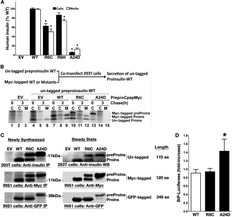
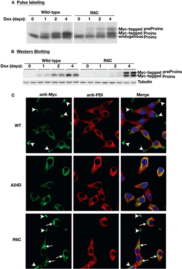
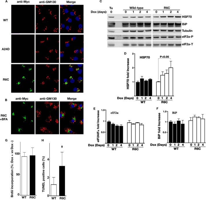
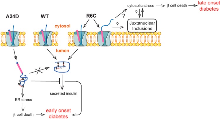
Among the defects in the early events of insulin biosynthesis, proinsulin misfolding and endoplasmic reticulum (ER) stress have drawn increasing attention as causes of β cell failure. However, no studies have yet addressed potential defects at the cytosolic entry point of preproinsulin into the secretory pathway. Here, we provide the first evidence that inefficient translocation of preproinsulin (caused by loss of a positive charge in the n region of its signal sequence) contributes to β cell failure and diabetes. Specifically, we find that, after targeting to the ER membrane, preproinsulin signal peptide (SP) mutants associated with autosomal dominant late-onset diabetes fail to be fully translocated across the ER membrane. The newly synthesized, untranslocated preproinsulin remains strongly associated with the ER membrane, exposing its proinsulin moiety to the cytosol. Rather than accumulating in the ER and inducing ER stress, untranslocated preproinsulin accumulates in a juxtanuclear compartment distinct from the Golgi complex, induces the expression of heat shock protein 70 (HSP70), and promotes β cell death. Restoring an N-terminal positive charge to the mutant preproinsulin SP significantly improves the translocation defect. These findings not only reveal a novel molecular pathogenesis of β cell failure and diabetes but also provide the first evidence of the physiological and pathological significance of the SP n region positive charge of secretory proteins.

Keywords: Cytosolic Protein Accumulation; Diabetes; Insulin Synthesis; Mutant; Preproinsulin; Proinsulin; Protein Translocation; β Cell.


Figures

FIGURE 1.
FIGURE 1.
The early events of insulin biosynthesis and defects in processing of preproinsulin to proinsulin caused by R6C or R6H. A, human preproinsulin has two methionines in its SP and six cysteines in its proinsulin moiety (top panel). 293T cells transfected with plasmids encoding preproinsulin WT or mutants were labeled with either [35S]Met/Cys or [35S]Met for 10 min. The cell lysates were immunoprecipitated with anti-insulin and analyzed using NuPage under reducing conditions. Pure [35S]Met labels only unprocessed human preproinsulin (preProins), whereas [35S]Met/Cys labels both human preproinsulin and proinsulin (Proins). Con, control. B, 293T cells transfected with empty vector (EV) or plasmids encoding preproinsulin WT or mutants were labeled with [35S]Met/Cys for 10 min followed by 0- or 90-min chase. Cell lysates (C) and chase media (M) were analyzed as in A. C, the newly synthesized preproinsulin WT and mutants from transfected 293T cells were labeled with [35S]Met/Cys for 10 min, immunoprecipitated, and analyzed using Tris-Tricine urea SDS-PAGE under non-reducing and reducing conditions. The asterisk denotes oxidized uncleaved A24D. D, human islets preincubated with 5.5 or 25.5 mm glucose for 20 h were labeled with either [35S]Met/Cys or [35S]Met for 10 min. The islet lysates were immunoprecipitated with anti-insulin and analyzed as in A. E, parallel groups of human islets of D were labeled with [35S]Met/Cys for 10 min, lysed, immunoprecipitated, and analyzed as C. Newly synthesized A24D from transfected 293T cells were used as a molecular weight control of uncleaved preproinsulin. F, quantification data of uncleaved preproinsulin in human islets and transfected 293T cells from at least three independent experiments shown in A–E.
FIGURE 2.
FIGURE 2.
Preproinsulin R6C and R6H are targeted to and strongly associated with the ER membrane but exhibit impaired translocation. A and B, effect of R6C on SRP-dependent protein targeting and translocation in vitro. Preproinsulin WT and mutants were synthesized by in vitro translation in the presence of [35S]Met. The ability of exogenously added SRP and SRP receptor to target preproinsulins to salt-washed and trypsin-digested ER microsomes (TKRM) was assayed by cleavage of the SP upon successful translocation (A) and by protection of the translocated protein from PK digestion (B), as described under “Experimental Procedures.” The translocation efficiency of the mutant proteins was normalized to that of preproinsulin WT, whose translocation at saturating SRP receptor concentration was set to be 100%. C, preproinsulin (preProins) R6C is targeted to the microsomes in an SRP-dependent manner. In vitro synthesis, targeting, and translocation of preproinsulin WT and R6C were carried out as in A and B in the presence and absence of exogenously added SRP and rough microsomes (RM). The reactions were loaded on a 0.5 m sucrose cushion, and microsomal membranes were sedimented by ultracentrifugation as described under “Experimental Procedures.” S, supernatant; P, pellet; Proins, proinsulin. D, quantification of data from two independent experiments shown in C. E, 293T cells coexpressing preproinsulin WT or mutants with a separate CMV promoter-driven GFP expressed in the cytosol were labeled with [35S]Cys/Met for 15 min and then incubated with or without 0.01% digitonin in PBS for 10 min to selectively permeabilize the plasma membrane. The cells transfected with empty vector (EV) served as controls. The membrane-bound and luminal proteins were sedimented at 14,000 rpm for 10 min. In digitonin-treated cells, although the majority of cytosolic GFP was liberated into supernatant, both cleaved and uncleaved preproinsulin remained in the pellet. IP, immunoprecipitation. F, 293T cells expressing preproinsulin R6C and A24D were labeled with [35S]Cys/Met for 20 min and subjected to the carbonate extraction as described under “Experimental Procedures.” Although proinsulin derived from R6C was mostly recovered in the supernatant (S) along with BiP, the majority of uncleaved preproinsulin R6C and A24D remained in the membrane pellet (P) along with calnexin, suggesting that uncleaved preproinsulin mutants are strongly associated with the ER membrane. T, total lysate.
FIGURE 3.
FIGURE 3.
The proinsulin moiety of preproinsulin R6C is exposed to the cytosol. A, 293T cells expressing Myc-tagged preproinsulin (preProins) R6C and A24D were subjected to PK digestion in the presence or absence of digitonin (DIG) or Triton X-100 (TRX), followed by anti-Myc Western blotting as described under “Experimental Procedures.” Unprocessed R6C was sensitive to PK digestion in partially permeabilized cells, indicating that its proinsulin (Proins) moiety was exposed to the cytosol. B, two engineered human preproinsulins with N-linked glycosylation sites were created, as indicated in the top panel. The newly synthesized preproinsulin WT and R6C labeled with either [35S]Met or [35S]Cys/Met were treated with or without PNGaseF before SDS-PAGE. Although proinsulin derived from WT or R6C was glycosylated (glyco-proins, lanes 3, 7, 9, and 11), unprocessed R6C labeled by pure [35S]Met (lanes 5 and 6) failed to acquire an N-linked glycan, indicating that unprocessed R6C was not delivered to the ER lumen.
FIGURE 4.
FIGURE 4.
The positive charge in the n region and the charge gradient flanking the h region of the SP play a critical role in the efficient targeting and translocation of preproinsulin. A, 293T cells transfected with empty vector (EV), human preproinsulin (preProins) WT, R6C, or M5R/R6C were labeled with [35S]Cys/Met for 15 min and analyzed by SDS-PAGE. Proins, proinsulin. B, the homology of human and rodent insulin gene 2 (Ins2) preproinsulin signal sequences. C, INS1 cells expressing Myc-tagged human (Hu) preproinsulin WT or R6C or mouse (Mo) Ins2 WT or R6C were examined by anti-Myc Western blotting (WB). Mouse Ins2 R6C shows a more severe translocation defect than human R6C. D, quantification data of uncleaved preproinsulin from at least three independent experiments shown in C. E, INS1 cells expressing Myc-tagged human preproinsulin WT or mutants were labeled with [35S]Met/Cys for 10 min. The newly synthesized preproinsulin was immunoprecipitated (IP) with anti-Myc, followed by SDS-PAGE. F, Myc-tagged human preproinsulin WT or mutants in transfected INS1 cells were examined by anti-Myc Western blotting. R6C/D20R significantly exacerbated the defect of R6C. G, quantification data of uncleaved preproinsulin from at least three independent experiments shown in F.
FIGURE 5.
FIGURE 5.
R6C and R6H fail to produce a normal amount of insulin but do not affect coexpressed preproinsulin WT nor induce ER stress. A, INS1 cells were transfected with empty vector (EV), human preproinsulin WT or mutants. At 48 h post-transfection, the human insulin in the lysates (black bars) and media (white bars) were measured using human insulin-specific RIA normalized to human insulin mRNA. Results shown are mean ± S.D. from three independent experiments. *, p < 0.05 compared with preproinsulin WT. B, 293T cells were cotransfected with untagged preproinsulin (preProins) WT, with empty vector (EV), or Myc-tagged preproinsulin WT, or mutants. At 48 h post-transfection, the cells were labeled for 15 min, followed by a 0- or 3-h chase. Cell lysates (C) and chase media (M) were immunoprecipitated with anti-insulin and analyzed under reducing conditions. Unlike A24D, which blocked the secretion of coexpressed WT, R6C did not affect the expression, translocation, and secretion of coexpressed WT. Proins, proinsulin. C, the plasmids encoding human preproinsulin WT, R6C, or A24D with or without a Myc or GFP tag was transfected into 293T or INS1 cells as indicated. The cells transfected with empty vector (EV) served as controls. After 48 h post-transfection, the expression and SP cleavage of untagged or tagged preproinsulin were examined and compared by pulse labeling (newly synthesized) and Western blotting (steady state) using anti-insulin, anti-Myc, or anti-GFP antibodies, as indicated. Unlike untagged and Myc-tagged preproinsulin R6C, there was no detectable defect in GFP-tagged R6C. IP, immunoprecipitation; aa, amino acids. D, BiP promoter activities in transfected pancreatic β cells were evaluated as described under “Experimental Procedures.” Cells expressing preproinsulin WT were served as a control. Results were expressed as mean ± S.D. from at least three independent experiments. *, p < 0.05 compared with preproinsulin WT.
FIGURE 6.
FIGURE 6.
Untranslocated R6C accumulates in a juxtanuclear region in pancreatic β cells. A, INS1 cells inducibly expressing Myc-tagged mouse preproinsulin (preProins) WT or mutants were established as described under “Experimental Procedures.” After Dox induction for the indicated days, the newly synthesized endogenous proinsulin (Proins), Myc-tagged preproinsulin WT, and R6C of inducible cell lines were labeled with [35S]Met/Cys for 15 min, immunoprecipitated with anti-insulin, and analyzed under reducing conditions. B, the steady state of Myc-tagged preproinsulin WT and R6C from the same sets of cells as in A was detected by anti-Myc Western blotting. C, after 4 days of Dox induction, INS1-inducible cell lines expressing Myc-tagged mouse preproinsulin WT, R6C, or A24D were pretreated with cycloheximide for 1 h before being fully permeabilized with 0.5% Triton X-100 and immunostained with anti-Myc (green) and anti-PDI (an ER marker, red) antibodies. Nuclei were counterstained with DAPI. In most cells expressing Myc-tagged preproinsulin WT (top row), anti-Myc immunoreactable molecules presented as a punctate, insulin granule-like pattern (arrowheads) that was distinct from PDI. For R6C (bottom row), two major intracellular pools were observed. One did indeed concentrate in distal tips (arrowheads), whereas another accumulated in a juxtanuclear region (arrows), and neither pool overlapped with PDI. A24D lost the granule pattern and largely overlapped with PDI.
FIGURE 7.
FIGURE 7.
Intracellular accumulation of untranslocated R6C induces cytosolic stress and promotes β cell death. A, the plasma membranes of similar sets of INA1-inducible cell lines as in Fig. 6C were selectively permeabilized by 0.01% digitonin and immunoblotted with anti-Myc (green) and anti-GM130 (a Golgi marker, red) antibodies. Nuclei were counterstained with DAPI. Unlike in Fig. 6C, anti-Myc immunoreactable molecules were detected only in the cells expressing Myc-tagged R6C, indicating that the proinsulin moiety of untranslocated R6C was exposed to the cytosol. Such molecules appeared to accumulate in a juxtanuclear region close to the Golgi marker GM130. B, parallel wells of the cells expressing Myc-tagged R6C as in A were pretreated with 5 μm brefeldin A (BFA) for 30 min to disrupt the Golgi structure before being partially permeabilized and immunoblotted as in A. The juxtanuclear accumulation persisted even after the Golgi architecture was disrupted by brefeldin A. C, the expression of HSP70, BiP, total eIF2a (eIF2a-T), and phosphorylated eIF2α (eIF2α-P) in INS1 inducible cell lines were examined by Western blotting after Dox induction the indicated days. The INS1 cells treated with 10 μm tunicamycin for 6 h served as controls. D–F, quantification data of HSP70, eIF2α-P, and BiP from two to four independent experiments shown C. G, BrdU incorporation of INS1-inducible cell lines after 4 days of Dox induction of Myc-tagged WT or R6C expression was measured as described under “Experimental Procedures.” H, cell death of the same sets of cells as in G was detected using TUNEL as described under “Experimental Procedures.” A total of ∼4500 cells expressing WT or R6C were counted from three independent experiments. The cells in parallel wells treated without Dox were used as controls. Results are shown as means + S.D. from three independent experiments. *, p < 0.05 compared with Myc-tagged preproinsulin WT.
FIGURE 8.
FIGURE 8.
A proposed model of β cell failure and diabetes caused by the defects in the earliest events of insulin biosynthesis. Two distinct underlying mechanisms associated with the defects occurring at the ER membrane can lead to β cell failure and diabetes. For preproinsulin A24D, impaired SP cleavage causes preproinsulin misfolding in the ER. Misfolded A24D not only induces ER stress but also blocks coexpressed proinsulin WT exit from the ER, causing a decrease of insulin production from proinsulin WT, leading to early-onset diabetes. For R6C, loss of the positive charge in the preproinsulin SP n region results in a misorientation of about 50% newly synthesized R6C molecules when their signal sequences interact with the Sec61 translocon. The misoriented R6C fails to be translocated into the ER lumen, accumulates in the cytosol and juxtanuclear compartment, induces cytosolic stress, and leads to β cell death and late-onset diabetes.

Similar articles

Cited by

References

    1. Dodson G, Steiner D. (1998) The role of assembly in insulin's biosynthesis. Curr. Opin. Struct. Biol. 8, 189–194 - PubMed
    1. Patzelt C., Labrecque A. D., Duguid J. R., Carroll R. J., Keim P. S., Heinrikson R. L., Steiner D. F. (1978) Detection and kinetic behavior of preproinsulin in pancreatic islets. Proc. Natl. Acad. Sci. U.S.A. 75, 1260–1264 - PMC - PubMed
    1. Wolin S. L., Walter P. (1993) Discrete nascent chain lengths are required for the insertion of presecretory proteins into microsomal membranes. J. Cell Biol. 121, 1211–1219 - PMC - PubMed
    1. Okun M. M., Shields D. (1992) Translocation of preproinsulin across the endoplasmic reticulum membrane. The relationship between nascent polypeptide size and extent of signal recognition particle-mediated inhibition of protein synthesis. J. Biol. Chem. 267, 11476–11482 - PubMed
    1. Liu M., Li Y., Cavener D, Arvan P. (2005) Proinsulin disulfide maturation and misfolding in the endoplasmic reticulum. J. Biol. Chem. 280, 13209–13212 - PMC - PubMed

Publication types