. 2014 Jun 20;289(25):17634-46.
doi: 10.1074/jbc.M114.552794. Epub 2014 Apr 25.

Interaction of branch migration translocases with the Holliday junction-resolving enzyme and their implications in Holliday junction resolution

Affiliations

Interaction of branch migration translocases with the Holliday junction-resolving enzyme and their implications in Holliday junction resolution

Cristina Cañas et al. J Biol Chem. .

Abstract

Double-strand break repair involves the formation of Holliday junction (HJ) structures that need to be resolved to promote correct replication and chromosomal segregation. The molecular mechanisms of HJ branch migration and/or resolution are poorly characterized in Firmicutes. Genetic evidence suggested that the absence of the RuvAB branch migration translocase and the RecU HJ resolvase is synthetically lethal in Bacillus subtilis, whereas a recU recG mutant was viable. In vitro RecU, which is restricted to bacteria of the Firmicutes phylum, binds HJs with high affinity. In this work we found that RecU does not bind simultaneously with RecG to a HJ. RuvB by interacting with RecU bound to the central region of HJ DNA, loses its nonspecific association with DNA, and re-localizes with RecU to form a ternary complex. RecU cannot stimulate the ATPase or branch migration activity of RuvB. The presence of RuvB·ATPγS greatly stimulates RecU-mediated HJ resolution, but the addition of ATP or RuvA abolishes this stimulatory effect. A RecU·HJ·RuvAB complex might be formed. RecU does not increase the RuvAB activities but slightly inhibits them.

Keywords: Atomic Force Microscopy (AFM); DNA Enzyme; DNA Helicase; DNA Repair; DNA-Protein Interaction; Genetic Recombination; Holliday Junction; RecG; RecU; RuvABC.


Figures

FIGURE 1.
FIGURE 1.
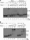
Detection of the RecU·RuvB·HJ complex. A, 0.4 nm γ-32P-labeled HJ-J3 DNA was preincubated with RuvB (40 nm) as indicated for 5 min at 37 °C in buffer B containing 15 mm CaCl2 and 1 mm ATP, then increasing concentrations of RecU or RecUR31A mutant, which is impaired in RuvB interaction, were added (10, 20, 40, 60 nm), and the samples were further incubated for 15 min at 37 °C. B, HJ-J3 was preincubated with RuvB (40 nm, lanes 2 and 7–10) or increasing RecU concentrations (5–100 nm, lanes 3–6) for 5 min at 37 °C in buffer B containing 15 mm CaCl2 and 1 mm ATPγS. As indicated, increasing concentrations of RecU (5–100 nm, lanes 7–10) were added, and all samples were further incubated for 15 min at 37 °C. Complexes were visualized using 6% PAGE and autoradiography. + and − indicate the presence and absence of protein, respectively. FD, free DNA; UD, RecU·DNA complexes; UBD, RecU·RuvB·DNA complexes. The amounts of RecU·DNA and RecU·RuvB·DNA complexes are given as percentages with respect of total DNA and are the average values obtained from >3 independent experiments (the results given stand within a 5% S.E.).
FIGURE 2.
FIGURE 2.
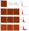
RecU recruits RuvB at the junction point. A, generation and visualization by AFM of the χ-structure. Induction of XerC recombinase promotes intramolecular recombination between cer sites (yellow) in pSD115, and the intermediate in this reaction is a χ-structure. HincII digestion (H) renders a long linear HJ with four arms of different length. The length of each arm is indicated. B–D, selected AFM images of the χ-structure incubated with RecU (20 nm, B), RuvB (10 nm, C), or both (D) in buffer containing 1 mm MgCl2 and 1 mm ATPγS for 10 min at 37 °C. White arrows denote protein-DNA complexes. When the χ-structure DNA is incubated with both RecU and RuvB, two type of complexes are observed (denoted by UBI and UBII). Frequency distributions of height of the RuvB-χ-structure (in C) and frequency distributions of height at the crossover point in the rest of conditions as well as the adjustment to a normal distribution were made with the program Origin 8.0. Scale bar = 500 nm.
FIGURE 3.
FIGURE 3.
Detection of the RecU·RuvA·HJ, RecU·RuvB·HJ and RecU·RuvA·RuvB·HJ complexes. A, high RecU concentrations favored the appearance of a quaternary complex. γ-32P-labeled HJ-jbm6 DNA (0.4 nm) was incubated with different combinations of RecUE36A (from 20 to 80 nm), RuvB (40 nm), and RuvA (80 nm) as indicated for 10 min at 37 °C in buffer B containing 5 mm MgCl2 and 1 mm ATPγS. Then anti-RecU (lanes 14–19) or 1× PBS was added (lanes 1–13), and incubation was continued for 10 min at 37 °C. Before electrophoresis, complexes were fixed by the addition of glutaraldehyde to a final concentration of 0.1% and 10 min incubation at 37 °C. B, antibody supershift experiments suggest the existence of ternary (RecU·RuvA·HJ and RecU·RuvB·HJ) and quaternary (RecU·RuvA·RuvB·HJ) complexes. γ-32P-Labeled HJ-jbm6 DNA was incubated with different combinations of RecUE36A (40 nm), RuvB (40 nm), and RuvA (80 nm) and then the indicated antibodies. In the lanes where no antibody was added, 1× PBS was added. + and − indicate the presence absence of protein, respectively. FD, free DNA; UD, RecU·DNA complexes; AD, RuvA·DNA complexes; AUD, RecU·RuvA·DNA complexes; ABD, RuvA· RuvB·DNA complexes; UBD, RecU·RuvB·DNA complexes; UABD, RecU·RuvA·RuvB·DNA complexes. The amounts of the different complexes observed are indicated as percentages with respect of total DNA and are the average values obtained from >3 independent experiments (the results given stand within a 5% S.E.). In lanes were an antibody was added, discrete complexes cannot be quantified because they were mostly shifted by the antibody.
FIGURE 4.
FIGURE 4.
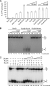
ATP or RuvA inhibits the stimulation of the cleavage promoted by RuvB. A, cleavage of the different arms of synthetic HJ-J3 by the RecU·RuvB complex. HJ-J3 labeled in the indicated arm was incubated with RecU (100 nm) and RuvB (40 nm) in buffer B containing 10 mm MgCl2 and 1 mm ATPγS for 30 min at 37 °C. 1*, 2*, 3*, and 4* denote the strand labeled in each condition. The reactions were analyzed by 15% denaturing PAGE and autoradiography, and the sites of DNA cleavage are shown at the right figure, which only shows the core of HJ-J3. The cleavage site detected by RecU alone is depicted with a black arrow. The new cleavage sites, detected in the presence of the RuvB·RecU complex, are shown by gray arrows. B, γ-32P-labeled HJs (HJ-J3 and HJ-J1, labeled in arm 1) were preincubated with RuvB (40 nm) for 5 min at 37 °C in buffer B containing 10 mm MgCl2 and 1 mm ATP or ATPγS as indicated, then RecU (100 nm) was added, and the reaction was continued for 30 min. Then reactions were deproteinized and analyzed by 6% native PAGE and autoradiography. C, the resolution of HJ-J3 by RecUY80A (300 nm) and RuvB (40 nm) is shown. The reactions were analyzed after deproteination by 6% PAGE and autoradiography. D, HJ-J3 and HJ-J1 were preincubated with RuvB (40 nm) in buffer B containing 10 mm MgCl2 and ATPγS, and then RecU (100 nm) and RuvA (100 nm) were added as indicated, and the reaction mixtures were further incubated for 30 min at 37 °C. Resolution was observed after deproteinization by 6% native PAGE and autoradiography. The amounts of cleavage on the given strand and resolution are indicated as percentages with respect of total DNA and are the average values obtained from >3 independent experiments (the results given stand within a 5% S.E.).
FIGURE 5.
FIGURE 5.
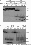
RecU does not stimulate RuvB activities. A, ATPase assays of RuvB in the presence of RecU and/or RuvA. RuvB (20 nm) was premixed on ice with 25 nm RuvA and/or 50–150 nm RecUE36A in the presence of HJ-J3 (100 μm in nucleotides) as effector DNA. Reactions were initiated by the addition of 1 mm ATP (mixed with [γ-32P]ATP, ratio 1*:100,000) and incubated for 15 min at 37 °C. Then reactions were stopped and analyzed as described under “Experimental Procedures.” The first two bars correspond to assays having only RecUE36A (150 nm) or only RuvA (25 nm). The −DNA, RuvB bar corresponds to assays where ATPase activity of RuvB in the absence of HJ DNA was assayed. B, branch migration activity by RuvB requires RuvA. γ-32P-labeled HJ-J3 DNA (0.4 nm) was incubated with 40 nm RuvB alone or in the presence of 12.5 nm RuvA or 100 nm RecUE36A in buffer B containing 10 mm MgCl2 and increasing ATP concentrations (0.1, 0.2, 0.5, and 1 mm) for 30 min at 37 °C. Reactions were deproteinized, and unwound products were analyzed by 6% PAGE and autoradiography. C, branch migration activity of RuvB in the presence of RuvA and RecU. γ-32P-labeled HJ-J3 DNA (0.4 nm) was incubated with 40 nm RuvB alone or in the presence of 12.5 nm RuvA and/or increasing amounts of RecUE36A (10, 50, 100, and 200 nm) as indicated in buffer B containing 10 mm MgCl2 and 1 mm ATP for 30 min at 37 °C, and the products of unwinding were analyzed by 6% PAGE and autoradiography. The amounts of unwound products are indicated as percentages with respect of total DNA and are the average values obtained from >3 independent experiments (the results given stand within a 5% S.E.).
FIGURE 6.
FIGURE 6.
Binding and unwinding of HJ-J3 by RecG. A, band shift assay showing the binding of RecG to HJ-J3 (lanes 1–8) and its unwinding (lanes 9–15). In lanes 1–8 doubling amounts of purified RecG (3.75–240 nm) were incubated with 0.4 nm γ-32P-labeled HJ-J3 in buffer B containing 5 mm EDTA for 15 min at 37 °C, put on ice, and subjected to 6% PAGE. In lanes 9–15, HJ-J3 DNA was incubated with the same amounts of RecG in buffer containing 5 mm MgCl2 and 5 mm ATP for 30 min at 37 °C followed by deproteinization. B, HJ-J3 unwinding as a function of time and ATP concentration. In lanes 2–7, HJ-J3 DNA was incubated for 30 min at 37 °C with 30 nm RecG in buffer B containing 5 mm MgCl2 and increasing concentrations of ATP as indicated. In lanes 9–15, HJ-J3 was incubated for a variable time at 37 °C with 30 nm RecG in buffer containing 5 mm MgCl2 and 5 mm ATP followed by deproteinization. FD, free HJ DNA; GD, RecG-DNA complexes. The amounts of GD complexes formed and unwound products (flayed substrates and the free oligonucleotide) are indicated as percentages with respect to total DNA and are the average values obtained from >3 independent experiments (the results given stand within a 5% S.E.).
FIGURE 7.
FIGURE 7.
RecU-mediated HJ cleavage is not stimulated by RecG. γ-32P-labeled HJ-J1 or HJ-J3 DNA (0.4 nm) was preincubated with RecG (30 or 60 nm) or RuvB (40 nm) in buffer B plus 10 mm MgCl2 and 1 mm ATPγS. Then RecU (100 nm) was added, and reactions were further incubated for 30 min at 37 °C before termination by stop-mix addition and analysis by 6% PAGE. In lanes 2 and 7, HJ-J1 and HJ-J3, respectively, were incubated with RecU alone for 30 min at 37 °C. The amount of resolution is indicated as percentage with respect of total DNA and is the average value obtained from >3 independent experiments (the results given stand within a 5% S.E.).
FIGURE 8.
FIGURE 8.
Binding and unwinding of HJ-J3 by RecG in the presence of RecU. A, RecU inhibits the helicase activity of RecG. In lanes 2–7, γ-32P-labeled HJ-J3 DNA (0.4 nm) was incubated only with one of the two proteins as indicated for 20 min at 37 °C; in lanes 7–12, HJ-J3 was incubated with the indicated amounts of RecUE36A for 5 min at 37 °C in buffer B containing 5 mm MgCl2 and 5 mm ATP. Then the indicated amounts of RecG were added, and reactions were continued for 20 min at 37 °C. After incubation, loading buffer was added. Samples separated on 6% PAGE allowed the observation of both, the binding of the proteins to the DNA, and the products of unwinding. B, RecU seems to displace RecG from HJ DNA. In lanes 9–16, 0.4 nm γ-32P-labeled HJ-J3 was incubated with the indicated amounts of RecG for 10 min at 37 °C in buffer B containing 5 mm EDTA and 5 mm ATP. Then the indicated amounts of RecUE36A were added and reactions continued for 10 min at 37 °C followed by 6% PAGE. In lanes 2–8, HJ-J3 was incubated with only 1 of the 2 proteins for 10 min at 37 °C under the same conditions. FD, free HJ DNA; GD, RecG-DNA complexes; UD, RecU-DNA complexes. The amounts of UD and GD complexes formed and unwound products are indicated as percentages with respect of total DNA and are the average values obtained from >3 independent experiments (the results given stand within a 5% S.E.).
FIGURE 9.
FIGURE 9.
Dissociation of RecG and RecU from HJ-J3. A, band-shift analysis of the dissociation of RecG and RecU from HJ-J3 in the presence of 5 mm EDTA and 1 mm ATPγS. γ-32P-labeled HJ-J3 DNA (0.4 nm, labeled in arm 1) was incubated with RecUE36A or RecG as indicated (4× KD app) for 15 min at 37 °C in a volume of 120 μl. Then an aliquot of 20 μl was taken and electrophoresed (lanes 2 and 9). A 50-fold excess of cold HJ-J3 was added to the rest, and 20-μl aliquots were taken at the indicated times and immediately subjected to 6% PAGE (lanes 3–7). B, stability of the RecUE36A-HJ-J3 and RecG-HJ-J3 complexes in the presence of 5 mm MgCl2 and 1 mm ATPγS. Reactions contained RecUE36A or RecG (4x KD app) and 0.4 nm HJ-J3, and after 15-min incubation, competitor DNA was added, and aliquots were taken at timed intervals. The amounts of RecU-DNA and RecG-DNA complexes formed are indicated as percentages with respect of total DNA and are the average values obtained from >3 independent experiments (the results given stand within a 5% S.E.). FD, free HJ DNA; GD, RecG-DNA complexes; UD, RecU-DNA complexes.

Similar articles

Cited by

References

    1. Holliday R. (1964) The induction of mitotic recombination by mitomycin C in Ustilago and Saccharomyces. Genetics 50, 323–335 - PMC - PubMed
    1. Mitchell A. H., West S. C. (1994) Hexameric rings of Escherichia coli RuvB protein. Cooperative assembly, processivity, and ATPase activity. J. Mol. Biol. 243, 208–215 - PubMed
    1. Fogg J. M., Schofield M. J., Déclais A. C., Lilley D. M. (2000) Yeast resolving enzyme CCE1 makes sequential cleavages in DNA junctions within the lifetime of the complex. Biochemistry 39, 4082–4089 - PubMed
    1. Ayora S., Carrasco B., Cárdenas P. P., César C. E., Cañas C., Yadav T., Marchisone C., Alonso J. C. (2011) Double-strand break repair in bacteria: a view from Bacillus subtilis. FEMS Microbiol. Rev. 35, 1055–1081 - PubMed
    1. Aravind L., Makarova K. S., Koonin E. V. (2000) Survey and summary: holliday junction resolvases and related nucleases. Identification of new families, phyletic distribution, and evolutionary trajectories. Nucleic Acids Res. 28, 3417–3432 - PMC - PubMed

Publication types

MeSH terms

LinkOut - more resources