. 2014 Jun 13;289(24):17268-79.
doi: 10.1074/jbc.M114.570390. Epub 2014 Apr 26.

Protein phosphatase 2A (PP2A) regulates low density lipoprotein uptake through regulating sterol response element-binding protein-2 (SREBP-2) DNA binding

Affiliations

Protein phosphatase 2A (PP2A) regulates low density lipoprotein uptake through regulating sterol response element-binding protein-2 (SREBP-2) DNA binding

Lyndi M Rice et al. J Biol Chem. .

Abstract

LDL-cholesterol (LDL-C) uptake by Ldlr is regulated at the transcriptional level by the cleavage-dependent activation of membrane-associated sterol response element-binding protein (SREBP-2). Activated SREBP-2 translocates to the nucleus, where it binds to an LDLR promoter sterol response element (SRE), increasing LDLR gene expression and LDL-C uptake. SREBP-2 cleavage and translocation steps are well established. Several SREBP-2 phosphorylation sites have been mapped and functionally characterized. The phosphatases dephosphorylating these sites remain elusive. The phosphatase(s) regulating SREBP-2 represents a novel pharmacological target for treating hypercholesterolemia. Here we show that protein phosphatase 2A (PP2A) promotes SREBP-2 LDLR promoter binding in response to cholesterol depletion. No binding to an LDLR SRE was observed in the presence of the HMG-CoA reductase inhibitor, lovastatin, when PP2A activity was inhibited by okadaic acid or depleted by siRNA methods. SREBP-2 cleavage and nuclear translocation were not affected by loss of PP2A. PP2A activity was required for SREBP-2 DNA binding. In response to cholesterol depletion, PP2A directly interacted with SREBP-2 and altered its phosphorylation state, causing an increase in SREBP-2 binding to an LDLR SRE site. Increased binding resulted in induced LDLR gene expression and increased LDL uptake. We conclude that PP2A activity regulates cholesterol homeostasis and LDL-C uptake.

Keywords: Cholesterol; DNA; Lipid; Lipoprotein; Low Density Lipoprotein (LDL); Metabolism; SREBP; Transcription.


Figures

FIGURE 1.
FIGURE 1.
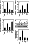
Okadaic acid inhibits lovastatin-induced gene expression. HepG2 cells were grown as described under “Experimental Procedures,” in the absence or presence of lovastatin (Lov), okadaic acid (OA), or lovastatin and okadaic acid (Lov OA) for 24 h. The PP2A inhibitor okadaic acid (10 nm) was added for the final 8 h, mRNA expression was determined using qRT-PCR, and protein level was determined by Western analysis using polyclonal antibodies. A, relative level of LDLR expression. B, relative level of SREBP-2 expression. C, relative level of HMGCR expression. D, Ldlr protein levels. E, relative densitometry units calculated using Ldlr protein levels in D. **, p < 0.001. Error bars, S.E.
FIGURE 2.
FIGURE 2.
siRNA directed against PP2A inhibits lovastatin-induced gene expression. HepG2 cells were grown as described under “Experimental Procedures,” in the absence (−Lov) or presence (+Lov) of lovastatin. Cells were treated with 100 nm non-targeting siRNA (Csi) or siRNA directed against the α catalytic subunit of PP2A (PPP2CAsi) at 48 and 24 h prior to lovastatin treatment. 75 μm lovastatin was added for 24 h, and cells were harvested. mRNA expression was determined using qRT-PCR. A, relative expression of PPP2CA. B, protein level of Ppp2ca. C, relative level of LDLR expression. D, relative level of ACC1 expression. E, relative level of FAS1 expression. F, relative level of PPARγ expression. G, Ldlr protein levels. H, relative densitometry units calculated using Ldlr protein levels in G. **, p < 0.001. GAPDH, glyceraldehyde phosphate dehydrogenase, loading control. Error bars, S.E.
FIGURE 3.
FIGURE 3.
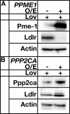
Altering the methylation status of Ppp2ca results in decreased SREBP-2 activity. HepG2 cells were transiently transfected with overexpression plasmids pCMV-PPME1 or pCMV-PPP2CA and were grown in the presence of 75 μm lovastatin for 24 h. Cells were harvested, and protein levels were determined by Western analysis. A, protein levels of Pme-1 and Ldlr. B, protein levels of Ppp2ca and Ldlr. **, p < 0.001. Actin, loading control.
FIGURE 4.
FIGURE 4.
Loss of Ppp2ca does not affect SREBP-2 cleavage and nuclear translocation. HepG2 cells were grown in the absence (Con) or presence of lovastatin (Lov). Cells were treated with 100 nm non-targeting siRNA (Csi) or siRNA directed against the catalytic subunit of PP2A (PPP2CAsi) at 48 and 24 h prior to lovastatin treatment. 75 μm lovastatin was added for 24 h, and cells were harvested. Cells were lysed and centrifuged as described under “Experimental Procedures” in order to obtain nuclear fractions. Cell protein was resolved by SDS-PAGE, and proteins were detected using Western analysis. Lamin was used as a nuclear marker. Gapdh was used as a non-nuclear marker. SREBP-2af, cleaved SREBP-2. A, non-nuclear fraction. B, nuclear fraction.
FIGURE 5.
FIGURE 5.
GFP-SREBP-2 translocates into the nucleus in cells lacking PP2A. HepG2 cells were transfected with no siRNA (Control), control siRNA (Csi), or siRNA directed against the catalytic subunit of PP2A (PPP2CAsi), in the absence (−Lov) or presence (+Lov) of lovastatin. pEGFP-SREBP-2 was transfected into cells that were subsequently treated with 40 μm lovastatin or DMSO for 1 h, fixed, and mounted with DAPI-containing mounting buffer to visualize nuclei. The movement of GFP-SREBP-2 was examined by fluorescence microscopy (Leica DMI6000B).
FIGURE 6.
FIGURE 6.
LDL-C uptake is defective in cells lacking Ppp2ca. HepG2 cells were transfected with control siRNA (Csi) or siRNA directed against the catalytic subunit of PP2A (PPP2CAsi) and were incubated for 16 h in lipoprotein-deficient medium to induce the expression of Ldlr. Cells were treated with 60 μm lovastatin or DMSO for 6 h. LDL-C uptake was initiated by incubating cells with 5 μg/ml BODIPY-LDL. Uptake of BODIPY-LDL was measured after 30 min at 37 °C. Cells were fixed and mounted with fluorescent mounting medium containing DAPI for counterstaining. Intercellular BODIPY-LDL was visualized using fluorescence microscopy.
FIGURE 7.
FIGURE 7.
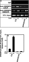
Loss of Ppp2ca causes a reduction in SREBP-2 LDLR SRE binding. Cells transfected with control siRNA (Csi) or siRNA directed against the catalytic subunit of PP2A (PPP2CAsi) were grown for 6 h in the absence (−Lov) or presence (+Lov) of lovastatin. Cells were then treated with 1% formaldehyde to cross-link SREBP-2 to DNA. The cross-linking reaction was terminated, and SREBP-2·DNA complexes were isolated using anti-SREBP-2 polyclonal antibodies. SREBP-2·DNA cross-links were disrupted using 5 m NaCl. A, amount of SREBP-2-bound DNA determined by PCR amplification and agarose gel electrophoresis. B, relative densitometry unit ratio of SREBP-2 SRE binding versus IgG binding. 10% of input, loading control; IgG, negative control. Error bars, S.E.
FIGURE 8.
FIGURE 8.
Ppp2ca is required for direct binding of SREBP-2 to a LDLR SRE. Nuclear extracts were prepared from HepG2 cells using a nuclear extract kit. WT and mutant probes were synthesized as single-stranded oligonucleotides. The sequences are given under “Experimental Procedures.” Nuclear extracts (NE) containing a biotinylated probe (Labeled Probe) were resolved on 6% non-denaturing polyacrylamide gels. Protein·DNA complexes were then transferred to Hybond-N+ nylon membranes and cross-linked. Detection of biotin-labeled DNA was performed using chemiluminescence and visualized by exposure to a charge-coupled device camera. For competition studies, a 10-fold molar excess of WT non-biotin labeled oligonucleotides (Unlabeled Probe) was used. For supershift experiments, SREBP-2 antibodies were added to the reaction mixture (SREBP-2 Ab). Gapdh antibodies were used as a negative control (Gapdh Ab). A, nuclear extracts were obtained from cells containing Ppp2ca in the absence of lovastatin (−Lov Ppp2ca). B, nuclear extracts were obtained from cells containing Ppp2ca in the presence of lovastatin (Lov Ppp2ca). C, nuclear extracts were obtained from cells lacking Ppp2ca in the absence of lovastatin (−Lov ppp2ca). D, nuclear extracts were obtained from cells lacking Ppp2ca in the presence of lovastatin (Lov ppp2ca).
FIGURE 9.
FIGURE 9.
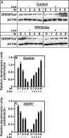
Loss of Ppp2ca has no effect on SREBP-2 half-life. HepG2 cells were transfected with 100 nm control siRNA (Con) or siRNA directed against the catalytic subunit of PP2A (PPP2CAsi) at −48 and −24 h in the absence (−Lov) and presence (+Lov) of lovastatin treatment. Cells were treated with 75 μm lovastatin for 24 h. Cells were treated with 5 μg/ml cyclohexamide for the indicated times, harvested, and stored at −80 °C until processing. Whole cell extracts were resolved using a 15% denaturing SDS-PAGE gel. Proteins were transferred to nitrocellulose and incubated with SREBP-2 antibodies. Protein was detected using ECL chemiluminescence. Actin, loading control. Error bars, S.E. A, Western analysis of SREBP-2 protein. B, densitometry of protein in control cells. C, densitometry of protein in ppp2a cells.
FIGURE 10.
FIGURE 10.
Ppp2ca interacts with SREBP-2. HepG2 cells were grown in the absence (−Lov) or presence (+Lov) of lovastatin. 75 μm lovastatin was added for 24 h. Whole cell lysates were obtained by sonication and centrifugation. Nuclear extracts were obtained as described under “Experimental Procedures.” Ppp2ca was immunoprecipitated (IP) using rat anti-Ppp2ca polyclonal antibodies. SREBP-2 was immunoprecipitated using SREBP-2 antibodies. Immunoprecipitated proteins were resolved by SDS-PAGE, and proteins were detected using Western analysis (WB). A, 10% of the whole cell extract used. B, immunoprecipitation of SREBP-2 using anti-Ppp2ca antibodies. C, immunoprecipitation of Ppp2ca using SREBP-2 antibodies. D, 10% of the nuclear extract used. E, immunoprecipitation of SREBP-2 using anti-Ppp2ca antibodies. F, immunoprecipitation of Ppp2ca using SREBP-2 antibodies. IgG, negative control.
FIGURE 11.
FIGURE 11.
Loss of Ppp2ca changes the phosphorylation state of SREBP-2. HepG2 cells were grown in the absence (−Lov) or presence (+Lov) of lovastatin. Cells were treated with 100 nm non-targeting siRNA (Csi) or siRNA directed against the catalytic subunit of PP2A (PPP2CAsi) at 48 and 24 h prior to lovastatin treatment. 75 μm lovastatin was added for 24 h. Cells were sonicated, cellular debris was pelleted by centrifugation, and cell lysates were collected. A, B, and C, phosphothreonine, phosphoserine, or phosphotyrosine proteins were used, immunoprecipitated (IP) by anti-phosphospecific polyclonal antibodies. Phosphorylated proteins were resolved by SDS-PAGE. The level of SREBP-2 phosphorylation was determined by Western analysis (WB) using SREBP-2 antibodies. The numbers represent relative densitometry units. All values are compared with their respective basal Csi value.

Similar articles

Cited by

References

    1. Maxfield F. R., Tabas I. (2005) Role of cholesterol and lipid and organization in disease. Nature 438, 612–621 - PubMed
    1. Sonnino S., Prinetti A. (2013) Membrane domains and the “lipid raft” concept. Curr. Med. Chem. 20, 4–21 - PubMed
    1. Wood W. G., Igbavboa U., Müller W. E., Eckert G. P. (2011) Cholesterol asymmetry in synaptic plasma membranes. J. Neurochem. 116, 684–689 - PMC - PubMed
    1. Elson E. L., Fried E., Dolbow J. E., Genin G. M. (2010) Phase separation in biological membranes. Integration of theory and experiment. Annu. Rev. Biophys. 39, 207–226 - PMC - PubMed
    1. Incardona J. P., Eaton S. (2000) Cholesterol in Signal Transduction. Curr. Opin. Cell Biol. 12, 193–203 - PubMed

Publication types